
|
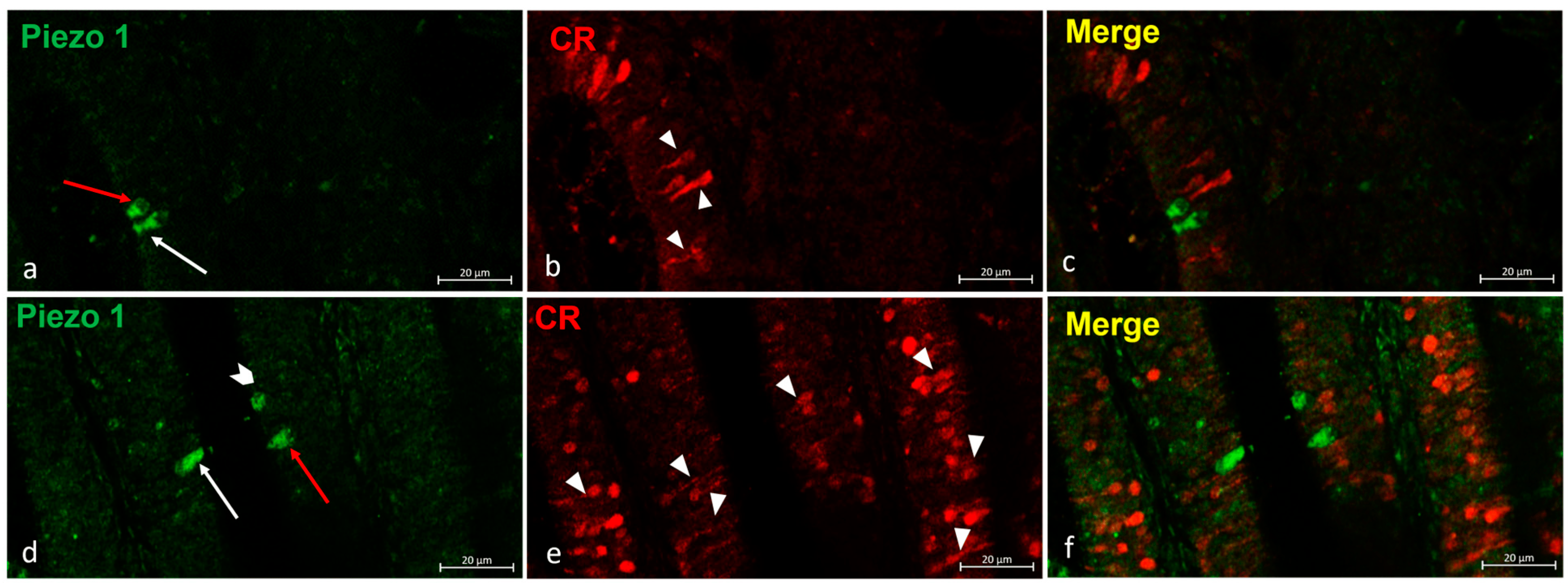
Fig. 2 (a–f) Zebrafish olfactory lamellae, dorsal view. We conducted an immunohistochemical detection (using the immunofluorescences method) of Piezo 1 in a colocalization view with Calretinin as a specific marker for the neuronal subpopulation cells. (a,d) The kappe cells (red arrows) and microvillous sensory cells (white arrows) were immunopositive to Piezo 1. (d) Piezo 1 was localized in crypt neurons (gallon arrow). (b,e) The ciliated sensory cells were immunolabeled to Calretinin (arrowhead). (c,f) A colocalization view that shows no overlap in the labelling. Magnification, 40×; scale bar, 20 µm.