
|
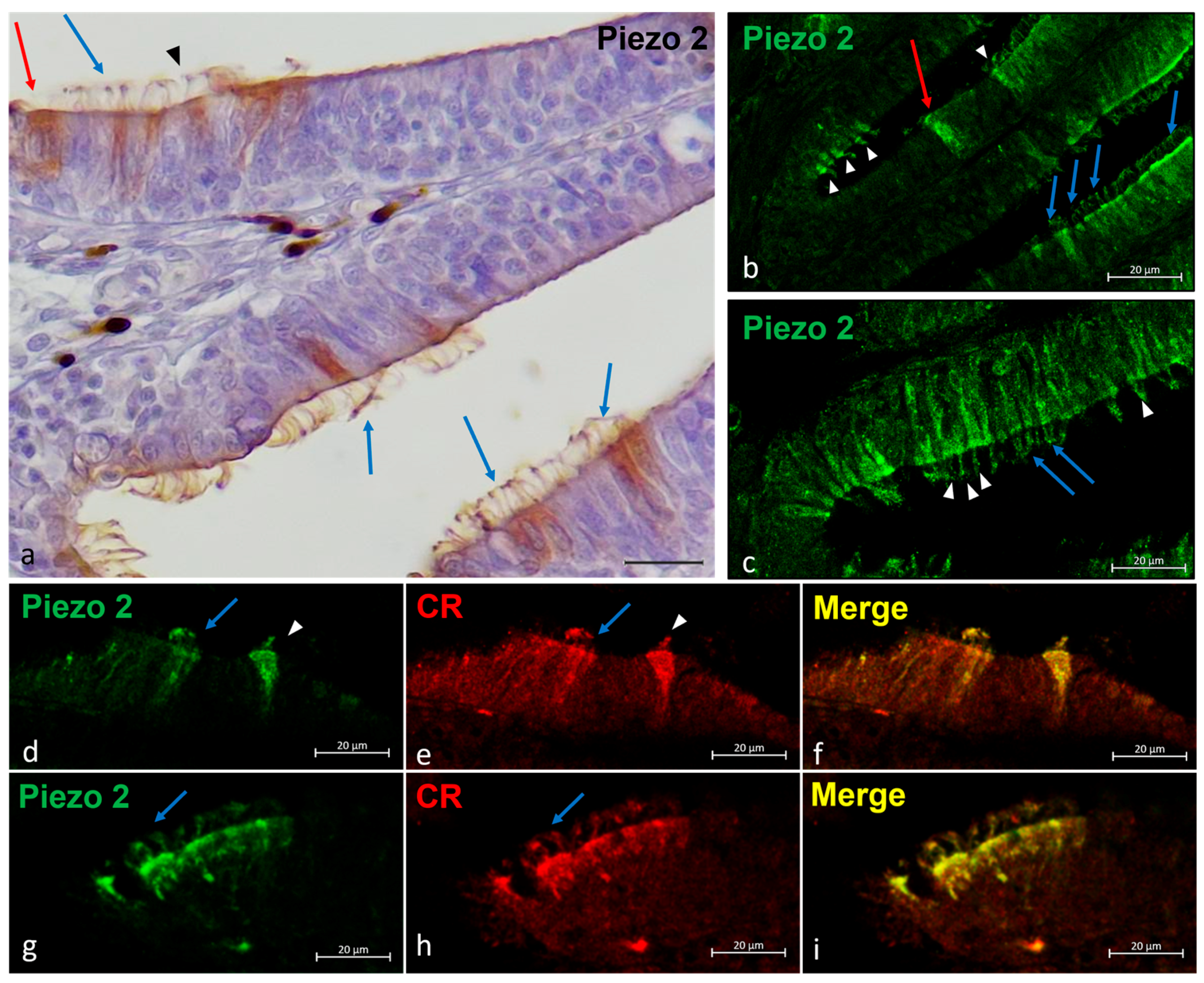
Fig. 4 Piezo 2 immunolocalizations in zebrafish olfactory lamellae, dorsal view. (a) The immunohistochemical detection (using a peroxidase method, Haematoxylin-stained) of Piezo 2 showing kappe neurons (red arrow), one-ciliated sensory cells (arrowhead), and multi-ciliated sensory cells (blue arrows). (b,c) The Piezo 2 immunolocalization showing kappe neurons (red arrow), one-ciliated sensory cells (arrowhead), and multi-ciliated sensory cells (blue arrows). (d,e,g,h) The immunohistochemical detection of Piezo 2 and Calretinin showing one-ciliated (arrowhead) and multi-ciliated (arrows) sensory cells. (f,i) A Piezo 2–Calretinin colocalization view. Magnification, 40×; scale bar, 20 µm