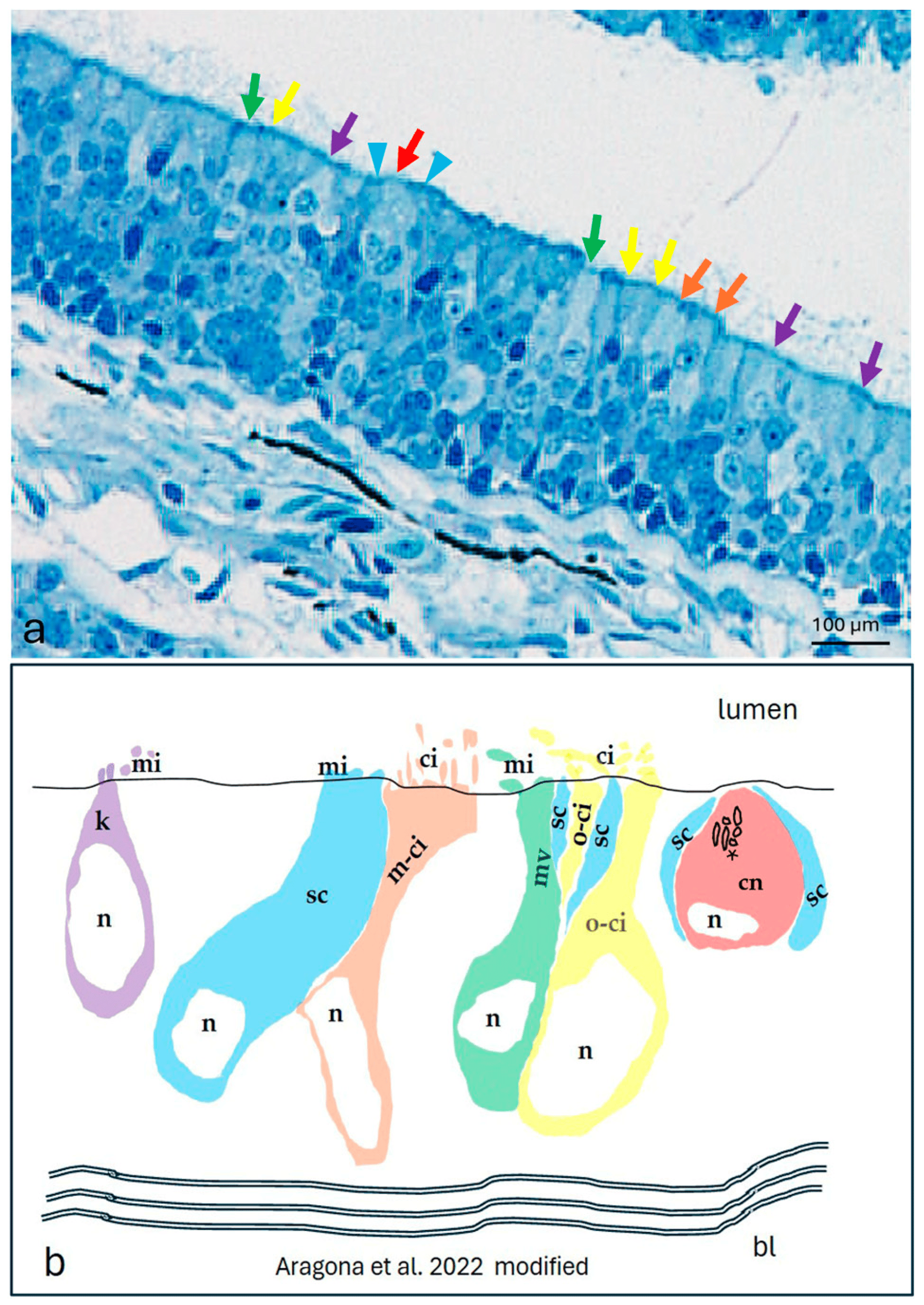
Fig. 3 The olfactory epithelium of an adult zebrafish. (a) Photomicrographs of the semithin section showing the kappe cells (purple arrows), one-ciliated ON (yellow arrows), multi-ciliated ON (orange arrows), microvillous ON (green arrows), and crypt neurons (red arrow) surrounded by special supporting cells (blue arrowheads). Toluidine blue; magnification, 20×. (b) A zebrafish epithelium olfactory neuron’s graphic representation modified using the transmission electron microscopy micrograph from our previous study [10]. Abbreviations: k, kappe cells; sc, supporting cells; m-ci, multi-ciliated ON; n, nucleus; o-ci, one-ciliated ON, ci, cilium; mv, microvillous ON; mi, microvillus; cn, crypt neurons, with several cilia within the crypt (asterisk), special supporting cells (sc), and basal lamina (bl)
                
                    
                        Image


                        Figure Caption


                        Acknowledgments


This image is the copyrighted work of the attributed author or publisher, and
ZFIN has permission only to display this image to its users.
Additional permissions should be obtained from the applicable author or publisher of the image.
      Full text @ Int. J. Mol. Sci.


        