
Image
|
Figure Caption
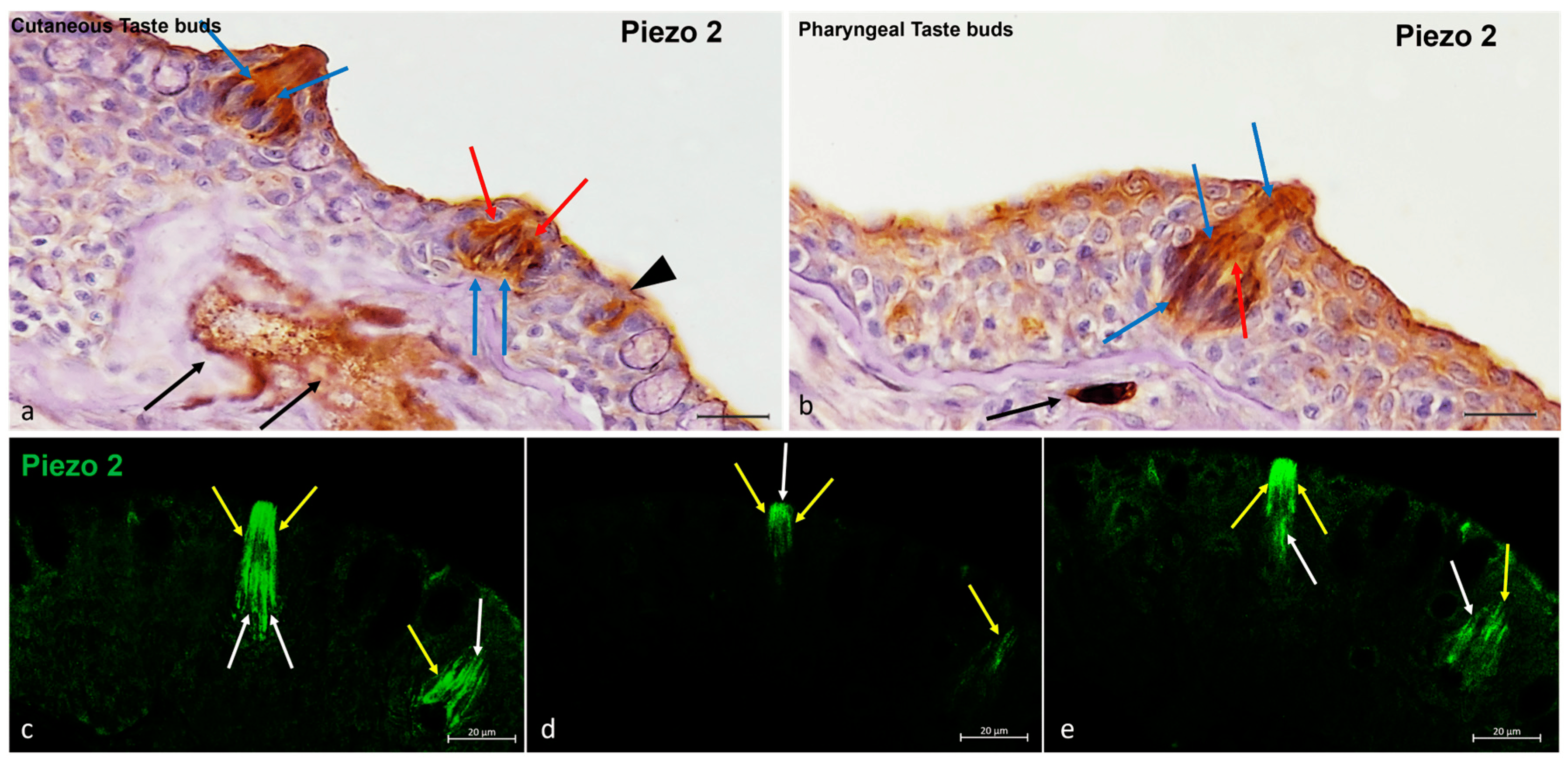
Fig. 6 Zebrafish taste buds, transversal view. (a,b) The immunohistochemical detection (using a peroxidase method, Haematoxylin-stained) of Piezo 2. The light cells (blue arrows), dark cells (red arrows), and nerve cells (black arrows) were Piezo 2-immunopositive. (a) Isolated chemosensory cells (black arrowhead) were immunoreactive to Piezo 2. (c–e) The multiple focal planes of the Piezo 2 immunohistochemical detections (using a fluorescence method). The light cells (white arrows) and dark cells (yellow arrows) were immunoreactive to Piezo 2. Magnification, 40×; scale bar, 20 µm.
Figure Data
Acknowledgments
This image is the copyrighted work of the attributed author or publisher, and
ZFIN has permission only to display this image to its users.
Additional permissions should be obtained from the applicable author or publisher of the image.
Full text @ Int. J. Mol. Sci.