
- Title
-
Arginine Vasotocin Directly Regulates Spermatogenesis in Adult Zebrafish (Danio rerio) Testes
- Authors
- Zanardini, M., Zhang, W., Habibi, H.R.
- Source
- Full text @ Int. J. Mol. Sci.
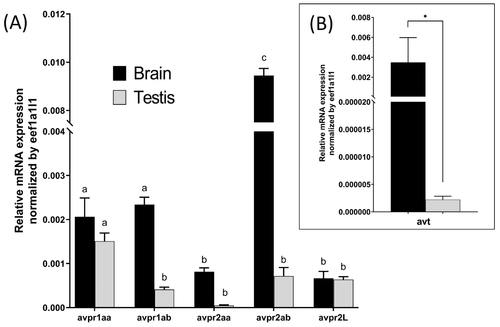
Relative transcript abundance of avt and Avp receptor subtypes in the testes and brain of zebrafish. Results show mRNA abundance of Avp receptor subtypes (avpr1aa, avpr1ab, avpr2aa, avpr2ab, avpr2l) (A) and vasotocin (avt) (B) in the adult zebrafish testis compared to the male brain. Columns represent basal expression levels from non-treated tissues (n = 5). Values were normalized with respect to eef1a1l1. Values with dissimilar superscripts are significantly different using a two-way ANOVA. Asterisk represents significant differences in transcript abundance of avt in the brain vs. testis, analyzed by the Mann–Whitney test. |
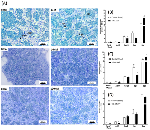
Dose-related effects of AVT on spermatogenesis. (A) Isolated contralateral testes were incubated separately, one as a control, the other with increasing concentrations of AVT. The arrangement of the figure reflects the way this experiment was conducted. The control for each concentration was from the same fish and different for different concentrations of AVT. Histological sections of zebrafish testes incubated ex vivo with or without (Basal) AVT for 7 days (A). Quantified cell types from testes incubated with 1 nM (B), 10 nM (C), and 100 nM (D) of AVT. The quantified results demonstrate the relative numbers of spermatogonia type A undifferentiated (Aund* + Aund), type A differentiated (Adiff), type B (SpgB), Spermatocytes (Spc), and Spermatozoa (Spz) for each concentration. Each treatment was analyzed with respect to its own (individual) control (contralateral testis). Asterisks represent significant differences between treatment and control, analyzed by Student’s t-test (n = 8–10), p < 0.05. |
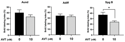
BrdU-labeling index of Aund, Adiff, and type B spermatogonia in basal conditions and in the presence of AVT (10 nM). BrdU was incorporated during the last 6 h of 7-day ex vivo testis culture. Bars show the mean ± SEM. Asterisks represent significant differences between treatment and control, analyzed by Student’s t-test (n = 5–7), p < 0.05. |
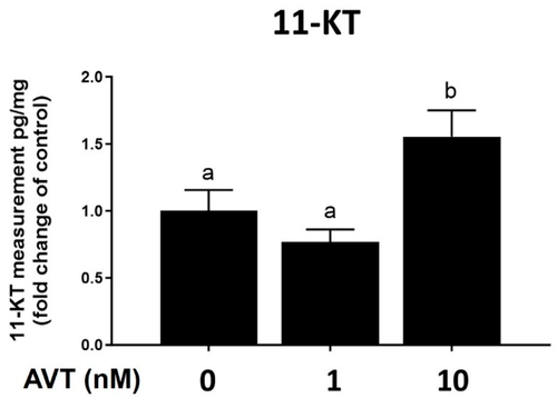
Quantification of 11-ketotestosterone in cultured testis medium after 7 days of treatment with 0, 1, and 10 nM vasotocin (AVT). Results are expressed as fold changes of their own (individual) control. Values are mean ± SEM of 6–8 replicate cultures. Different letters indicate significant differences between groups (one-way ANOVA followed by Tukey’s multiple comparison test, p < 0.05). |

The effect of 10 μM of flutamide (FLU), alone or in combination with 10 nM vasotocin (AVT), on (A) spermatogonia type Aund* + Aund, (B) Adiff, and (C) haploid spermatids and spermatozoa, following 7 days of ex vivo culture. Cells were counted in the control vs. treatment groups, and each treatment group was normalized against its control and expressed as fold change (mean ± SEM; n = 8–10). Values displaying different symbols are significantly different (one-way ANOVA followed by Tukey’s multiple comparison test, * p < 0.05, ** p < 0.01, *** p < 0.001). |
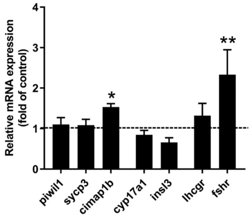
Relative transcript abundance of piwil1, sycp3, cimap1b (previously odf3b), cyp17a1, insl3, lhcgr, and fshr in testes sampled after 48 h of ex vivo treatment with 10 nM AVT. The dotted line represents the control group. Each treatment is presented as fold change to the control. The results were analyzed with respect to their own (individual) control using Student’s t-test (n = 8–10). Values displaying different symbols are significantly different compared to the control (* p < 0.05, ** p < 0.01). |