
|
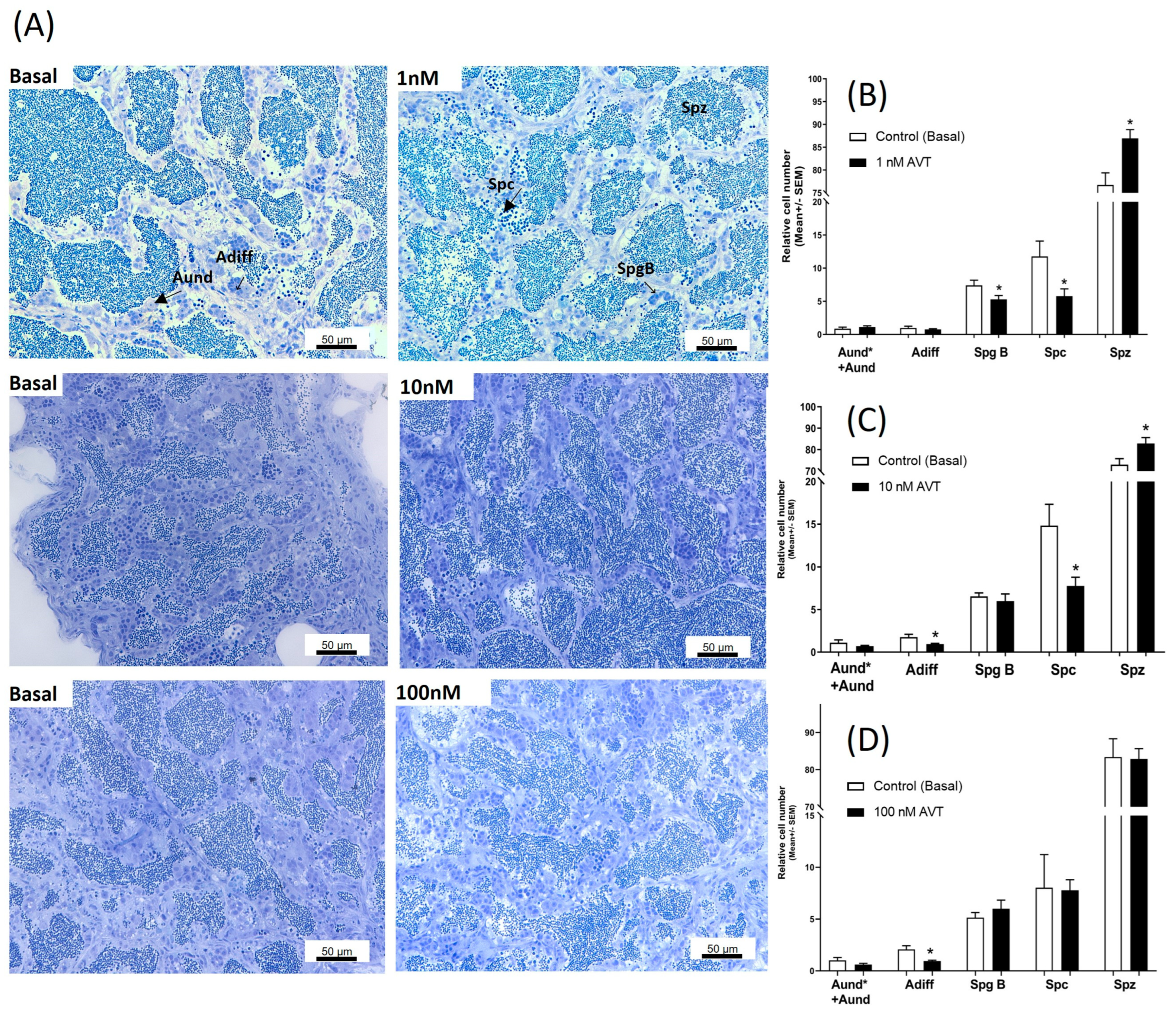
Fig. 2 Dose-related effects of AVT on spermatogenesis. (A) Isolated contralateral testes were incubated separately, one as a control, the other with increasing concentrations of AVT. The arrangement of the figure reflects the way this experiment was conducted. The control for each concentration was from the same fish and different for different concentrations of AVT. Histological sections of zebrafish testes incubated ex vivo with or without (Basal) AVT for 7 days (A). Quantified cell types from testes incubated with 1 nM (B), 10 nM (C), and 100 nM (D) of AVT. The quantified results demonstrate the relative numbers of spermatogonia type A undifferentiated (Aund* + Aund), type A differentiated (Adiff), type B (SpgB), Spermatocytes (Spc), and Spermatozoa (Spz) for each concentration. Each treatment was analyzed with respect to its own (individual) control (contralateral testis). Asterisks represent significant differences between treatment and control, analyzed by Student’s t-test (n = 8–10), p < 0.05.