FIGURE SUMMARY
Title

Shedding a Light on Dark Genes: A Comparative Expression Study of PRR12 Orthologues during Zebrafish Development

Authors
Muscò, A., Martini, D., Digregorio, M., Broccoli, V., Andreazzoli, M.
Source
Full text @ Genes (Basel)

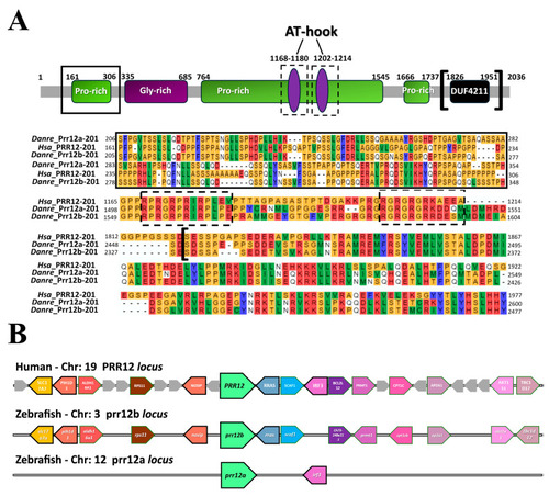
Multiple in silico approaches unveil conservative features between PRR12 and its orthologs in zebrafish. (A) Schematic representation of human PRR12 protein highlighting the main acknowledged domains (modified from Chowdhury et al. [7]), together with a multiple sequence alignment of the amino-terminal proline-rich (black box; 35% identity Prr12a-201 and Prr12b-201 vs. PRR12), AT-hook (black dashed-line boxes; AT-hook 1168–1180aa: 91.66% identity Prr12a-201 and Prr12b-201 vs. PRR12; AT-hook 1202–1214: 66.67% identity Prr12a-201 and Prr12b-201 vs. PRR12) and DUF4211 (black squared brackets; 60% identity Prr12a-201 and Prr12b-201 vs. PRR12) functional motifs of the human and zebrafish Prr12a-201 and Prr12b-201 proteins. Abbreviations: Hsa:Homo sapiens; Danre: Danio rerio. (B) Syntenic relationship between human PRR12 and zebrafish prr12a and prr12b loci. A total of six genes is conserved in prr12b surroundings compared to chromosome 19. A poor synteny in conversely evidenced for prr12a locus. Chr: chromosome.

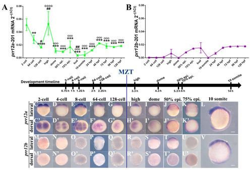
prr12a and prr12b display a different spatio-temporal expression pattern during zebrafish early development. (A,B) Real-time PCR profile of prr12a-201 (A) and prr12b-201 (B) at increasing developmental stages (epi.: epiboly; hpf: hours post fertilization). Ct values for each timepoint are normalized to rps11 gene. Asterisks (*) indicate statistical comparisons between each stage and 2 cell stage; hash marks (#) indicate statistical comparisons between each stage and 64 cell stage; Phi (Φ) indicate statistical comparison between each stage and 128 cell stage; degree symbol (°) indicate statistical comparison between each stage and high stage. Data are expressed as mean ± SD. ANOVA followed by Tukey post hoc test: ** p < 0.01; *** p < 0.001; **** p < 0.0001; # p < 0.05; ## p < 0.01; ΦΦΦΦ p < 0.0001; °°° p < 0.0001. n = 2 independent experiments. (CV) WISH using prr12a-201 anti-sense (CL) and prr12b-201 (MV) anti-sense probes at early developmental stages. Lateral (CL,MV) and dorsal (C’K’,M’U’) views of the hybridized embryos. Scale bars: 250 µm. Abbreviations: epi.: epiboly h: hours; MZT: maternal-to-zygotic transition.

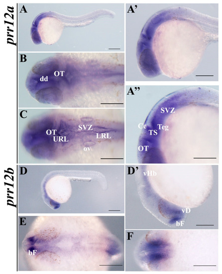
prr12a-201 and prr12b-201 are distributed differently in 24 hpf embryos. WISH analysis of prr12 expression at 24 hpf stage. (A,D) Lateral views of 24 hpf hybridized embryos and magnification of heads shown in (A’) and (D’). (A’’) Enlargement of prr12a-201-positive periventricular area. (B,C) forebrain and hindbrain dorsal views of prr12a hybridized embryos. (E,F) Dorsal and ventral views, respectively, of prr12b-201 hybridized embryos. Scale bars: 500 µm. Abbreviations: bF: basal forebrain; Ce: cerebellum; dd: dorsal diencephalon; LRL: lower rhombic lip; OT: optic tectum; ov: otic vesicle; SVZ: subventricular zone; TS: torus semicircularis; Teg: tegmentum; URL: upper rhombic lip; vHb: ventral hindbrain; vD: ventral diencephalon.

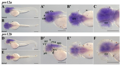
prr12 co-orthologs cover distinct domains of 48 hpf central nervous system. WISH evaluation of prr12 expression at 48 hpf. Scale bars: 500 µm. (A,D) lateral and (B,E) dorsal views of hybridized embryos. (A’,D’,B’,E’) represent the enlargements of the heads of the embryos shown in (A,D,B,E), respectively. (C,F) are the same embryos shown in (B’,E’), respectively, slightly tilted on a side and analyzed at higher magnifications to better show the hybridization signal in the eye. Abbreviations: DT: dorsal thalamus; H: hypothalamus; MO: medulla oblongata; P: pallium; PTd: dorsal part of posterior tuberculum; PTv: ventral part of posterior tuberculum; T: mesencephalic tegmentum; VT: ventral thalamus.

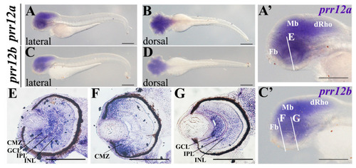
prr12a-201 and prr12b-201 territories converge in 72-hpf embryos. WISH (AD,A’,C’) and ISH on cryosectioned larval heads (EG). (A,C) lateral and (B,D) dorsal views of hybridized larvae. (A’,C’) enlargements of the heads of the embryos, respectively shown in (A,C). White lines indicate the section planes shown in (EG). Scale bars in (AD) and (A’C’): 500 µm; Scale bars in (E): 250 µm. Abbreviations: CMZ: ciliary marginal zone; dRho: dorsal rhombencephalon; Fb: forebrain; GCL: ganglion cell layer; INL: inner nuclear layer; IPL: inner plexiform layer; Mb: midbrain.

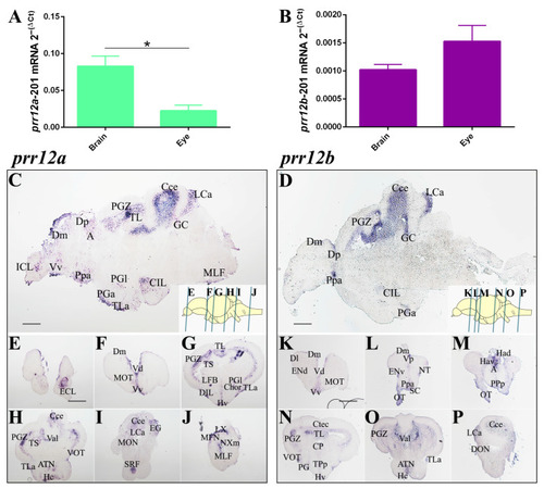
PRR12 zebrafish orthologs are expressed in adult brain and eye. (A,B) Relative quantification of prr12a-201 (A) and prr12b-201 (B) by RT-qPCR. Ct values are normalized to rps11 gene. Data are presented as mean ± SEM. Unpaired t test. * p < 0.05. n = 3 independent experiments. (CP) ISH on sagittal (C,D) and transversal (EP) adult brain cryosections. prr12a-201 and prr12b-201 mRNAs are found in proliferative niches and in nuclei variably associated with the olfactory, attentive, emotional and visual systems. Blue bars in the schematic depictions of the sagittal brain shown on the bottom right in (C,D) pictures, indicate the cutting planes of the cryosections shown in (EP). In (C,D), as well in the relative brain schemes, anterior is to the left and dorsal is to the top. Scale bars: 50 µm. Abbreviations: A: anterior thalamic nucleus; ATN: anterior tuberal nucleus; Cce: cerebellar corpus; CG: central gray; Chor: horizontal commissure; CIL/DIL: central/dorsal nucleus of the inferior lobe; Ctec: tectal commissure; CP: central posterior nucleus of dorsal hypothalamus; Dm/l/p: medial/lateral/posterior zone of dorsal telencephalic area; ECL/ICL: external/internal cellular layer of olfactory bulb; ENv: entopeduncular nucleus—ventral part; Hav/Had: ventral/dorsal habenular nucleus; Hc/d: caudal/dorsal zone of periventricular hypothalamus; Lca: caudal lobe of cerecellum; LFB: lateral olfactory tract; LX: vagal lobe; MFN: medial funicular nucleus; MLF: medial longitudinal fascicle; MOT: medial olfactory tract; NXM: vagal motor nucleus; PGZ: periventricular gray zone of optic tectum; TL: longitudinal torus; PGa/l: anterior/lateral preglomerular nucleus; Ppa/p/v: parvocellular preoptic nucleus-anterior/posterior/ventral part; SRF: superior reticular formation; TLa: lateral torus; TPp: periventricular nucleus of posterior tuberculum; TS: semicircular torus; Val: lateral division of valvula cerebelli; VOT/OT: ventrolateral/optic tract; Vv: ventral nucleus of ventral telencephalic area.

Acknowledgments
This image is the copyrighted work of the attributed author or publisher, and ZFIN has permission only to display this image to its users. Additional permissions should be obtained from the applicable author or publisher of the image. Full text @ Genes (Basel)