IMAGE

Figure 6

ID
ZDB-IMAGE-240429-129
Genes
Source
Figures for Muscò et al., 2024
Image
Figure Caption

Figure 6

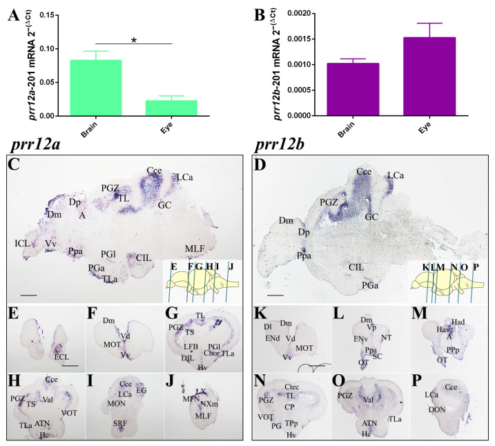
PRR12 zebrafish orthologs are expressed in adult brain and eye. (A,B) Relative quantification of prr12a-201 (A) and prr12b-201 (B) by RT-qPCR. Ct values are normalized to rps11 gene. Data are presented as mean ± SEM. Unpaired t test. * p < 0.05. n = 3 independent experiments. (CP) ISH on sagittal (C,D) and transversal (EP) adult brain cryosections. prr12a-201 and prr12b-201 mRNAs are found in proliferative niches and in nuclei variably associated with the olfactory, attentive, emotional and visual systems. Blue bars in the schematic depictions of the sagittal brain shown on the bottom right in (C,D) pictures, indicate the cutting planes of the cryosections shown in (EP). In (C,D), as well in the relative brain schemes, anterior is to the left and dorsal is to the top. Scale bars: 50 µm. Abbreviations: A: anterior thalamic nucleus; ATN: anterior tuberal nucleus; Cce: cerebellar corpus; CG: central gray; Chor: horizontal commissure; CIL/DIL: central/dorsal nucleus of the inferior lobe; Ctec: tectal commissure; CP: central posterior nucleus of dorsal hypothalamus; Dm/l/p: medial/lateral/posterior zone of dorsal telencephalic area; ECL/ICL: external/internal cellular layer of olfactory bulb; ENv: entopeduncular nucleus—ventral part; Hav/Had: ventral/dorsal habenular nucleus; Hc/d: caudal/dorsal zone of periventricular hypothalamus; Lca: caudal lobe of cerecellum; LFB: lateral olfactory tract; LX: vagal lobe; MFN: medial funicular nucleus; MLF: medial longitudinal fascicle; MOT: medial olfactory tract; NXM: vagal motor nucleus; PGZ: periventricular gray zone of optic tectum; TL: longitudinal torus; PGa/l: anterior/lateral preglomerular nucleus; Ppa/p/v: parvocellular preoptic nucleus-anterior/posterior/ventral part; SRF: superior reticular formation; TLa: lateral torus; TPp: periventricular nucleus of posterior tuberculum; TS: semicircular torus; Val: lateral division of valvula cerebelli; VOT/OT: ventrolateral/optic tract; Vv: ventral nucleus of ventral telencephalic area.

Figure Data
Acknowledgments
This image is the copyrighted work of the attributed author or publisher, and ZFIN has permission only to display this image to its users. Additional permissions should be obtained from the applicable author or publisher of the image. Full text @ Genes (Basel)