|
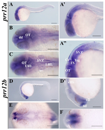
prr12a-201 and prr12b-201 are distributed differently in 24 hpf embryos. WISH analysis of prr12 expression at 24 hpf stage. (A,D) Lateral views of 24 hpf hybridized embryos and magnification of heads shown in (A’) and (D’). (A’’) Enlargement of prr12a-201-positive periventricular area. (B,C) forebrain and hindbrain dorsal views of prr12a hybridized embryos. (E,F) Dorsal and ventral views, respectively, of prr12b-201 hybridized embryos. Scale bars: 500 µm. Abbreviations: bF: basal forebrain; Ce: cerebellum; dd: dorsal diencephalon; LRL: lower rhombic lip; OT: optic tectum; ov: otic vesicle; SVZ: subventricular zone; TS: torus semicircularis; Teg: tegmentum; URL: upper rhombic lip; vHb: ventral hindbrain; vD: ventral diencephalon.
|

