FIGURE SUMMARY
Title

MEndoB, a chimeric lysin featuring a novel domain architecture and superior activity for the treatment of staphylococcal infections

Authors
Roehrig, C., Huemer, M., Lorgé, D., Arn, F., Heinrich, N., Selvakumar, L., Gasser, L., Hauswirth, P., Chang, C.-.C., Schweizer, T.A., Eichenseher, F., Lehmann, S., Zinkernagel, A.S., Schmelcher, M.
Source
Full text @ MBio

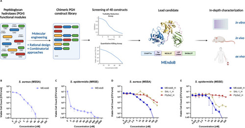
Selection of MEndoB from a library of chimeric staphylococcal PGHs. A chimeric PGH library of 48 constructs was cloned, expressed, and characterized (A). Constructs were screened for activity in PBS and human serum with two orthogonal assays (turbidity reduction assay and quantitative killing assay [qKA]) against S. aureus and S. epidermidis. The most active construct (MEndoB) was further tested in vitro, ex vivo, and in vivo. MEndoB comprises two EADs (N-terminal CHAP domain [blue] and the central M23 endopeptidase domain [yellow]), one CBD (C-terminal SH3b domain [green]), and two linkers (purple and pink) from different origins, shown in a 3D-modeled ribbon representation (ColabFold). Activity of MEndoB against S. aureus ATCC 12600 (B) and S. epidermidis ATCC 35984 (C) in human serum, as determined by qKAs. Activity of C-terminally His-tagged versions of MEndoB, SAL-1, and PlySs2 in human serum against S. aureus ATCC 12600 (D) and S. epidermidis ATCC 12228 (E), determined by qKAs. The limit of detection was 200 CFUs/mL (dotted line), and error bars represent standard error of the mean from four (B and C) and two (D and E) biological replicates. Figure was modified from reference 32 and created using biorender.com.

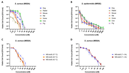
MEndoB kills different staphylococcal species in various animal sera, at elevated temperatures, and remains stable upon storage for 4 h at 37°C in human serum. MEndoB activity in qKAs at 37°C against S. aureus ATCC 12600 (A) and S. epidermidis ATCC 35984 (B) in seven different animal sera and at 37°C, 40°C, and 42°C against S. aureus ATCC 12600 in human serum (C). Data shown in panel C for 37°C corresponds to data from Fig. 1B. Activity of MEndoB after incubation in human serum for 4 h at 37°C was determined in qKAs at 37°C against S. aureus ATCC 12600 (D). The limit of detection was 200 CFUs/mL (dotted line), and error bars indicate the standard error of the mean from two (A and B) and three (C and D) biological replicates.

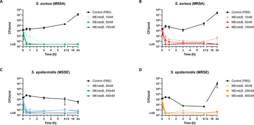
MEndoB effectively kills staphylococcal species in human whole blood. Time kill curves of different S. aureus and S. epidermidis strains in fresh human whole blood are shown. Human whole blood was inoculated with 104 CFUs/mL S. aureus ATCC 12600 (MSSA) (A), S. aureus USA300 JE2 (MRSA) (B), S. epidermidis 1457 (MSSE) (C), or S. epidermidis ATCC 35984 (MRSE) (D) and MEndoB or PBS was added at three different concentrations [10, 50, and 100 nM for (A and B) and 40, 200, and 400 nM for (C and D)]. Bacterial survival was evaluated over time (5 min, 30 min, 1 h, 3 h, 6 h, and 24 h) (A–D). The limit of detection was 27 CFUs/mL (dotted line), and error bars show the standard error of the mean from four healthy donors. 100 nM = 4.6 µg/mL.

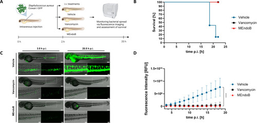
MEndoB rescues zebrafish larvae infected with S. aureus. Overview of the experimental procedure (A). Forty-eight-hour-old zebrafish larvae were injected with S. aureus Cowan I pCN56_GFPmut2 (2.5 × 10⁴ CFUs/fish) and treated with vehicle (PBS), vancomycin, or MEndoB 2 h post-infection. Survival of treated zebrafish larvae was monitored (n = 7) (B). Real-time GFP fluorescence images were overlayed with phase contrast microscopy images (C) and their intensity was quantified over time (D). Scale bar = 500 mm, scale bar of insets = 200 mm. Mean ± SD is shown (n = 7). ****P ≤ 0.0001, repeated measure two-way ANOVA.

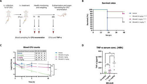
Treatment with MEndoB results in dose-dependent survival of up to 90% in a lethal mouse bacteremia model. Schematic representation of the experimental procedure (A). Survival rates of mice infected with S. aureus USA300 LAC strain treated with vehicle (PBS) or MEndoB at 0.1 mg/kg (gray), 1.0 mg/kg (pink), and 10.0 mg/kg (green) were monitored over 48 h post-treatment (n = 10 per group) (B). CFUs in the blood of treated animals were analyzed after 0, 1, 6, and 48 h (C). The limit of detection was 2.7 Log10 CFUs/mL (dotted line). The number of analyzed blood samples is shown in the table; only non-deceased animals with clinical scores permitting blood sampling were included in the analysis (no vehicle group beyond 6 h time point). TNF-α serum concentration in the blood of the mice infected with S. aureus USA300 LAC strain was analyzed 48 h post-treatment (D). For all graphs, error bars indicate the standard error of the mean. Statistical analysis performed for survival rates was a log-rank (Mantel-Cox) test (vehicle vs treatment groups) and an unpaired t-test at 48 h for blood CFU counts and TNF-α concentrations (****P < 0.0001, ***P ≤ 0.001, **P ≤ 0.01, *P ≤ 0.05, and ns > 0.05).

Acknowledgments
This image is the copyrighted work of the attributed author or publisher, and ZFIN has permission only to display this image to its users. Additional permissions should be obtained from the applicable author or publisher of the image. Full text @ MBio