
- Title
-
Aerobic exercise enhances mitochondrial homeostasis to counteract D-galactose-induced sarcopenia in zebrafish
- Authors
- Chen, Z.L., Guo, C., Zou, Y.Y., Feng, C., Yang, D.X., Sun, C.C., Wen, W., Jian, Z.J., Zhao, Z., Xiao, Q., Lan-Zheng, ., Peng, X.Y., Zhou, Z.Q., Tang, C.F.
- Source
- Full text @ Exp. Gerontol.
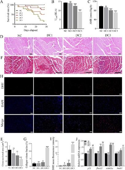
A dip in d-galactose concentrations causes sarcopenia in zebrafish. (A) Survival curves for each group of zebrafish maintained under different concentration gradients of D-galactose (d-gal) solution (0, 200, 400, 800, or 1600 mg/L). (B, C) Effects of different concentrations of d-gal solution on the critical swimming speed (Ucrit) and exercise oxygen consumption (AMR) of zebrafish. (D) Hematoxylin and eosin staining of skeletal muscles in each zebrafish group. Scale bar: 500 μm. (E) The cross-sectional area of skeletal muscles (μm2) in each zebrafish group. (F) Masson's trichome staining of zebrafish skeletal muscle tissues in each group. Blue and red illustrate stained collagen fibers and myofibers, respectively. Scale bar: 100 μm. (G) Fibrosis fraction (%) of zebrafish in each group. (H) Dihydroethidium (DHE) staining of zebrafish skeletal muscle tissues in each group. Red and blue show reactive oxygen species (ROS) and nuclear staining, respectively. Scale bar: 100 μm. (I) Fluorescence quantification value of ROS stains for each zebrafish group. (J) mRNA expression of senescence and muscle atrophy marker genes, such as p53, fbxo32, trim63a, and lmnb1, in skeletal muscles of each group of zebrafish. * represents d-gal-treated controls (DC) compared with normal controls (NC). * p < 0.05. *** p < 0.001. ns, not significant. |

Aerobic exercise counteracts the d-galactose-induced reductions in skeletal muscle function and mass. (A) Changes in the body size of zebrafish in each group after treatment. (B) Body weights of zebrafish in each group at the end of the 4-week treatment period. (C, D) Effects of aerobic exercise on the critical swimming speed (Ucrit) and exercise oxygen consumption (AMR) in D-galactose (d-gal)-induced aging zebrafish. (E) Hematoxylin and eosin staining of skeletal muscles in each group of zebrafish. Scale bar: 500 μm. (F) The cross-sectional area of skeletal muscle (μm2) in each zebrafish group. (G) Masson's trichome staining of zebrafish skeletal muscle tissue in each group. Blue and red show collagen fiber and myofiber staining, respectively. Scale bar: 100 μm. (H) Fibrosis fraction (%) of each zebrafish group. * represents d-gal-treated controls (DC) compared with normal controls (NC); # represents d-gal aerobic exercise (DA) compared with DC. #, * p < 0.05. ###, *** p < 0.001. ns, not significant. (For interpretation of the references to colour in this figure legend, the reader is referred to the web version of this article.) |
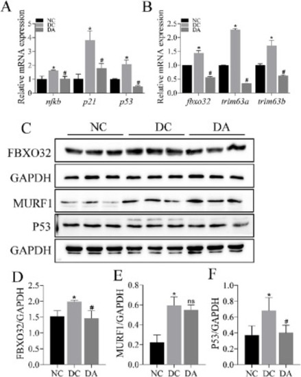
Aerobic exercise alleviates the d-galactose-induced skeletal muscle aging and damage. (A) mRNA expression of nfκb, p21, and p53 (skeletal muscle senescence-associated genes) in zebrafish. (B) mRNA expression of fbxo32, trim63a, and trim63b (skeletal muscle atrophy-related genes) in zebrafish. (C) Protein expression of skeletal muscle senescence and myasthenia-related genes in zebrafish. (Dsingle bondF) Protein expression results. * represents d-gal-treated controls (DC) compared with normal controls (NC); # represents d-gal aerobic exercise (DA) compared with DC. #, * p < 0.05. ns, not significant. |
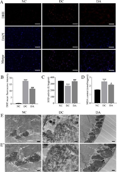
Aerobic exercise alleviates the d-galactose-induced oxidative stress and mitochondrial damage in skeletal muscles. (A) Dihydroethidium (DHE) staining of skeletal muscle tissues for each zebrafish group. Red and blue show reactive oxygen species (ROS) and nuclear staining, respectively. Scale bar: 100 μm. (B) Quantitative fluorescence values of ROS staining for each zebrafish group. (C) Effects of aerobic exercise on superoxide dismutase (SOD) activity in skeletal muscles of d-gal-induced senescent zebrafish. (D) Malondialdehyde (MDA) content in skeletal muscles of zebrafish. (E) Transmission electron micrographs of the skeletal muscles of zebrafish. Scale bar: 50 nm. (E') Magnification of local mitochondria. Scale bar: 34.5 nm. * represents d-gal-treated controls (DC) compared with normal controls (NC); # represents d-gal aerobic exercise (DA) compared with DC. # p < 0.05. ###, *** p < 0.001. (For interpretation of the references to colour in this figure legend, the reader is referred to the web version of this article.) |
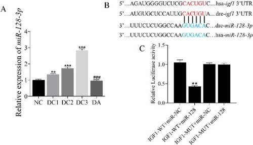
Aerobic exercise downregulates miR-128 and activates insulin-like growth factor-1 signaling in aging skeletal muscle. (A) Effect of different concentrations of d-galactose with aerobic exercise on miR-128 expression in zebrafish skeletal muscle. (B) An analysis of the human and zebrafish insulin-like growth factor-1 (IGF-1) sequence for a potential miR-128 binding site. The blue and red sections indicate the seed region of miR-128 and the binding site of the seed region to igf1 3′UTR, respectively. (C) Quantitative analysis of the dual-luciferase reporter assay. IGF-1-WT: synthesized wild-type IGF-1 3’UTR pmirGLO vector that contains the miR-128 predicted binding site; IGF-1-MUT: synthesized mutated IGF-1 3’UTR pmirGLO vector that contains the mutated miR-128 predicted binding site; miR-NC: negative control miRNA. * represents d-gal-treated controls (DC) compared with normal controls (NC); # represents d-gal aerobic exercise (DA) compared with DC. #, * p < 0.05. ## p < 0.01. ###, *** p < 0.001. (For interpretation of the references to colour in this figure legend, the reader is referred to the web version of this article.) |
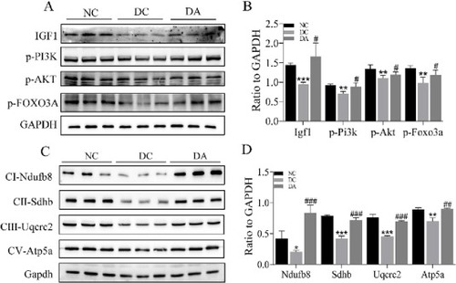
Aerobic exercise activates the insulin-like growth factor-1 signaling pathway in zebrafish skeletal muscle and enhances mitochondrial respiratory function. (A, B) Protein expression of Igf1 and phosphorylation of Pi3k, Akt, and Foxo3a are upregulated after aerobic exercise. (C, D) Expression of Ndufb8, Sdhb, Uqcrc2, and Atp5a, which are key proteins of the mitochondrial respiratory chain I-V, is significantly upregulated after aerobic exercise. * represents d-gal-treated controls (DC) compared with normal controls (NC); # represents d-gal aerobic exercise (DA) compared with DC. #, * p < 0.05. ##, ** p < 0.01. ###, *** p < 0.001. GAPDH, glyceraldehyde 3-phosphate dehydrogenase. |
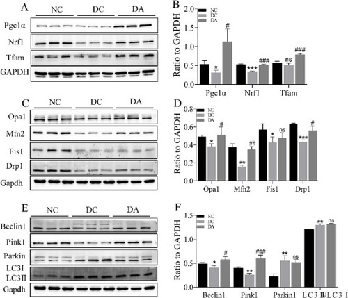
Aerobic exercise enhances mitochondrial homeostasis in aging skeletal muscle. (A, B) Protein expression results for mitochondrial biogenesis-related proteins in zebrafish skeletal muscles. (C, D) Protein expression results for mitochondrial dynamics-related protein expression in skeletal muscles of zebrafish. (E, F) Expression of mitochondrial autophagy-related proteins in skeletal muscles of zebrafish. * represents d-gal-treated controls (DC) compared with normal controls (NC); # represents d-gal aerobic exercise (DA) compared with DC. #, * p < 0.05. ##, ** p < 0.01. ###, *** p < 0.001. GAPDH, glyceraldehyde 3-phosphate dehydrogenase. |
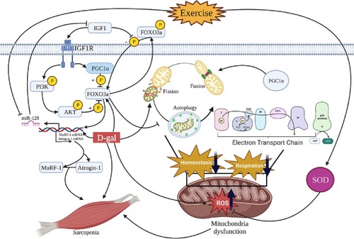
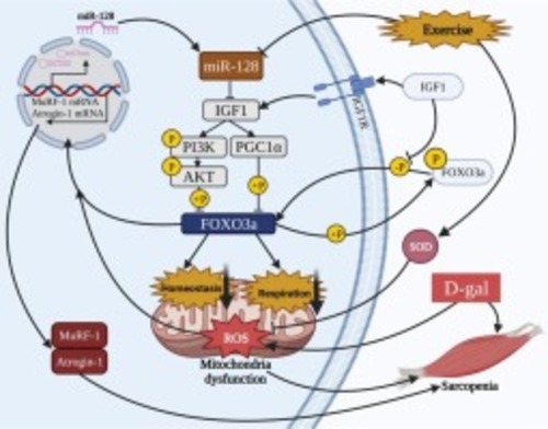
Putative mechanistic map of the mechanisms by which aerobic exercise resists sarcopenia through the miR-128/IGF-1 signaling pathway. d-galactose (d-gal) contributes to the development of sarcopenia by causing metabolic disturbances in vivo to produce large amounts of reactive oxygen species (ROS) and promote the expression of muscle atrophy-related factors MuRF1, Atrogin1, and miR-128. Aerobic exercise inhibits miR-128 expression, which activates the expression of its direct target, insulin-like growth factor-1 (IGF-1). IGF-1 may further promote phosphorylation of FOXO3a by activating PI3K/AKT and PGC1-α signaling pathways. These changes may improve mitochondrial function impaired by d-gal induction, thereby resisting sarcopenia. ATP, adenosine triphosphate; ADP, adenosine diphosphate; SOD, superoxide dismutase. |
|