
- Title
-
Leukemia-associated truncation of granulocyte colony-stimulating factor receptor impacts granulopoiesis throughout the life-course
- Authors
- Bulleeraz, V., Goy, M., Basheer, F., Liongue, C., Ward, A.C.
- Source
- Full text @ Front Immunol

ZFIN is incorporating published figure images and captions as part of an ongoing project. Figures from some publications have not yet been curated, or are not available for display because of copyright restrictions. PHENOTYPE:
|
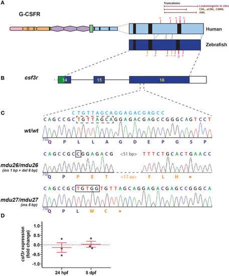
Generation of leukemia-derived G-CSFR truncation mutants. Schematic diagram of the G-CSFR showing the extracellular immunoglobulin domain (pink), cytokine receptor homology domain (orange) and fibronectin type III-like domains (purple), transmembrane region (green), and intracellular region (blue) containing Box 1-3 (black rectangles). The intracellular region is expanded to show tyrosine (Y) residues and a di-leucine motif (LL) in both human and zebrafish proteins, with the relative positions of truncation mutations found in various human leukemias or demonstrated to be leukemogenic in vitro shown above (A). Exons 14-16 of the zebrafish csf3r gene encoding the intracellular region presented as numbered boxes with connecting lines depicting introns (B). Sequence traces of the indicated part of csf3r showing homozygous wild-type (wt/wt) and mutant zebrafish, with nucleotide sequence above and encoded protein sequences below, in purple for native and orange for de novo sequence (C). The gRNA target site is shown above in blue, with deleted nucleotide sequences indicated with a brown dotted box and inserted sequences shown with black boxes. The csf3rmdu26 (mdu26) allele represents a complex 1 bp insertion/8 bp deletion and the csf3rmdu27 (mdu27) allele a 5 bp insertion both of which cause a frameshift resulting in translation from an alternative reading frame followed by a stop codon. Gene expression analysis of csf3r in pooled WT and mutant embryos at the indicated timepoints presented as fold-change (log2) relative to actb (D), showing mean and SEM (not significant, n=3). |

Effect of G-CSFR truncation mutation on primitive hematopoiesis. Wild-type (wt/wt), heterozygous (wt/mdu26) and homozygous (mdu26/mdu26) mutant csf3r embryos were subjected to WISH at 22 hpf with mpo (A–C), spi1b (E–G) and gata1a (I–K), with representative images shown. Individual embryos were assessed for the number of mpo + (D) or spi1b + (H) cells or the area of gata1a (L) staining, with mean and SEM in red and level of statistical significance indicated (***: p < 0.001, ns: not significant; n=20-30). EXPRESSION / LABELING:
PHENOTYPE:
|

Effect of G-CSFR truncation mutation on definitive hematopoiesis. Wild-type (wt/wt), heterozygous (wt/mdu26) and homozygous (mdu26/mdu26) mutant csf3r embryos were subjected to WISH at 5 dpf with mpo (A–C), mpeg1.1 (E–G), rag1 (I–K) and hbbe1.1 (M–O), with representative images shown. Individual embryos were assessed for the number of mpo+ (D) or mpeg1.1+ (H) cells or the area of staining for rag1 (L) or hbbe1.1 (P), with mean and SEM shown in red and level of statistical significance indicated (***: p < 0.001, ns: not significant; n=20-28). |

Effect of G-CSFR truncation mutation on adult hematopoiesis. (A–D). Adult blood cells from wild-type (wt/wt), heterozygous (wt/mdu26) and homozygous (mdu26/mdu26) mutant csf3r fish were subjected to Giemsa-staining (A–C), along with differential quantitation of the indicated blood cell populations for individual fish (D), with mean and SEM shown in red and level of statistical significance indicated (ns: not significant; n=4). Abbreviation: n: neutrophil. (E–P). Adult kidney cells from wild-type (wt/wt), heterozygous (wt/mdu27) and homozygous (mdu27/mdu27) mutant csf3r fish on a Tg(mpo::GFP) background were subjected to FACS analysis using SSC/FSC (E–G) and GFP fluorescence (I–K), along with quantitation of indicated cell populations (H) and GFP+ neutrophils (L), with sorted GFP+ cells subjected to Giemsa-staining (M–O), along with differential quantitation of relative differentiation (P). Panels (H, L, P) display results for individual fish, with mean and SEM shown in red and level of statistical significance indicated (*: p < 0.05, ns: not significant; n=4-9). im, immature; int; intermediate; m, mature. |
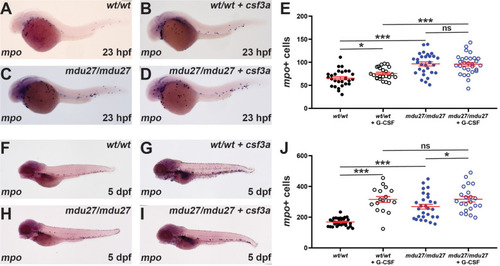
Effect of G-CSFR truncation mutation on emergency hematopoiesis. Wild-type (wt/wt) and homozygous (mdu27/mdu27) mutant csf3r embryos, either uninjected or injected with mRNA encoding G-CSF (+ csf3a) were subjected to WISH with mpo during primitive (A–D) and early definitive (F–I) hematopoiesis, with representative images shown. Individual embryos were assessed for the number of mpo+ cells for primitive (E) and definitive (J) hematopoiesis, with mean and SEM shown in red and level of statistical significance indicated (***: p < 0.001, *: p < 0.05, ns: not significant; n=17-30). EXPRESSION / LABELING:
PHENOTYPE:
|