FIGURE SUMMARY
Title

Mvda is required for zebrafish early development

Authors
Wong, W., Huang, Y., Wu, Z., Kong, Y., Luan, J., Zhang, Q., Pan, J., Yan, K., Zhang, Z.
Source
Full text @ Biol. Res.

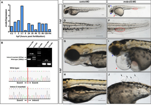
Aberrant mvda function causes developmental defects in zebrafish. A Quantitative real-time PCR (qRT-PCR) for twelve embryo development stages (0.2 hpf, 1 hpf, 2 hpf, 3.7 hpf, 6 hpf, 24 hpf, 30 hpf, 48 hpf, 72 hpf, 96 hpf, 120 hpf, and 144 hpf) demonstrates different expression patterns of mvda during embryonic development. B Effectiveness of mvda knockdown was confirmed by RT-PCR and sanger sequencing. The zebrafish mvda gene was targeted by specific morpholino antisense to prevent the proper splicing of exon 3 (E3I3-MO). Primers spanning mvda exon 1 (forward) and exon 4 (reverse) interrogate the presence of wild type (non-mutant) transcripts or those in which intron 3 has been inserted. RT-PCR of mvda transcript from control-MO and E3I3-MO injected embryos at 2-dpf, demonstrating insertion of intron 3. Sanger sequencing of both the wild-type band and the intron 3-inserted band validating the wild-type sequence and the intron 3-inserted sequence. CJ Gross morphology at 2-dpf and 4-dpf. Compared with the control group, knockdown of mvda presented hydrocephaly (E, blue arrowhead), eye defects (E, I, blue arrow), pericardial oedema (E, I, red arrow), blood accumulation in the caudal vein (E, F, red circled area), and skin defects (J, black arrows). Heartbeat and circulation in the caudal vein were visible in the control fish but were abnormal in mvda morphants (Additional file 2: Video S1, Additional file 3: Video S2). hpf, hours post fertilization; dpf, days post fertilization. Scale bars = 100 µm

EXPRESSION / LABELING:
Gene:
Fish:
Knockdown Reagent:
Anatomical Term:
Stage Range: 1-cell to Day 6

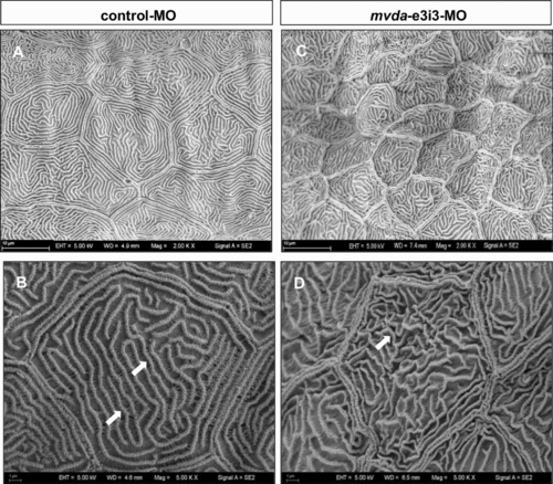
SEM of the skin surface in 4-dpf control fish and mvda morphants. A, B Control group revealed well-demarcated keratinocytes with regular arranged microridges (B, white arrows). C, Dmvda-e3i3-MO-injected zebrafish revealed keratinocytes with shrunken borders and microridges. The microridges showed tortuous architecture and irregular intervals (D, white arrow). dpf, days post fertilization. Scale bars = 10 µm (A, C) and 1 µm (B, D)

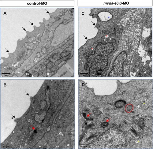
TEM of 4-dpf larvae injected with control or mvda morpholinos. A, B Keratinocytes of the control group revealed regular arranged spicules (black arrows) and cytoplasmic keratin filaments. C, D Keratinocytes of the mvda-e3i3-MO-injected zebrafish revealed disorganized spicules (black arrows), disordered cytoplasmic keratin filaments, distended rough endoplasmic reticulum (yellow asterisks), swollen mitochondria (red circle), autophagosomes and mitophagy (red arrowheads), vesicles (red asterisks), and rupture of cell–cell contacts (blue asterisk). Open arrowheads point to the basement membrane; e, epidermis; d, dermis; cs, collagenous stroma; dpf, days post fertilization. Scale bars = 1 µm (A, C) and 0.5 µm (B, D)

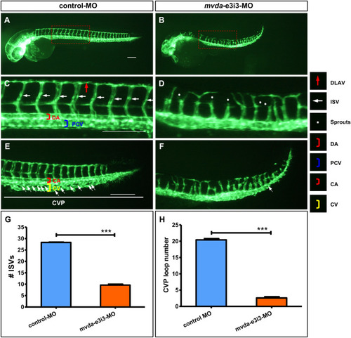
Morpholino knockdown of mvda impairs the trunk angiogenesis and CVP formation in zebrafish. AF Representative fluorescent images of Tg(fli1a:EGFP)y1 embryos at 2-dpf, with the vascular structures visualized by eGFP fluorescence. The boxed regions of A and B are shown at a higher magnification in C and D, respectively. C ISVs and DLAV showed regular development in the embryos injected with control-MO. D Compared with the control group, embryos injected with mvda-e3i3-MO presented a lower number of incomplete ISVs and ectopic sprouts (asterisks) of DA. E In control embryos, CVP was formed honeycomb-like structures at the tail around 2-dpf (white arrows). F In contrast, mvda knockdown resulted in specific defects in CVP formation. G Quantification of the number of complete ISVs shows a significant decrease in mvda morphants. H Quantification of loop formation at CVP shows a 7.8-fold decrease in mvda morphants. Columns, mean; bars, SEM (n = 10; unpaired student’s t-test) ***P < 0.0001. ISV intersegmental vessel, DLAV dorsal longitudinal anastomotic vessels, DA dorsal aorta, CVP caudal vein plexus, PCV posterior cardinal vein, CA caudal artery, CV caudal vein, dpf days post fertilization. Scale bars = 100 µm

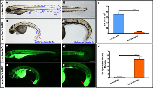
mvda deficiency induces fin-reduction phenotypes and tail-specific apoptosis. A normal fins, control-MO; B fin-reduction phenotypes after mvda-e3i3-MO injected; EF caudal fin is shown at higher magnification. Dashed lines indicate the morphology of fins. Control-MO-injected embryos and embryos injected with mvda-e3i3-MO were stained with acridine orange at 4-dpf. Apoptotic cells are visible as bright green spots, and less bright homogenous green or black staining is unspecific background staining. C, G Control-MO-injected zebrafish exhibited few or no apoptotic cells in whole organism. D, H In contrast, significantly increased staining was observed throughout the tail in zebrafish injected with mvda-e3i3-MO (red arrows). I Quantification of area at fin shows a 13.9-fold decrease in mvda morphants. Error bars, s.e.m.; ***P < 0.0001(n = 10; ANOVA). J Quantification of apoptosis particle number at tail shows a 25.8-fold increase in mvda morphants. Error bars, s.e.m.; ***P < 0.0001(n = 10; Student’s t test); A–H: lateral view, anterior, left. CF caudal fin, DF dorsal fin, PF pelvic fin, VF ventral fin, dpf days post fertilization. Scale bars = 100 µm

PHENOTYPE:
Fish:
Knockdown Reagent:
Observed In:
Stage: Day 4

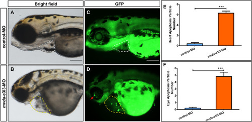
Morpholino knockdown of mvda induces potent apoptosis in heart and eyes. Control-MO-injected embryos and embryos injected with mvda-e3i3-MO were stained with acridine orange at 4-dpf. Apoptotic cells are visible as bright green spots, and less bright homogenous green staining is unspecific background staining. A, C Control-MO-injected zebrafish exhibited few or no apoptotic cells in heart and eyes. B, D In contrast, significantly increased staining was observed throughout the heart and eyes in zebrafish injected with mvda-e3i3-MO (circled areas and red arrow). E Quantification of apoptosis particle number at heart shows a 15.8-fold increase in mvda morphants. F Quantification of apoptosis particle number at eyes shows a 24-fold increase in mvda morphants. Error bars, s.e.m.; ***P < 0.0001(n = 10; Student’s t test); A–D: lateral view, anterior, left. dpf days post fertilization. Scale bars = 100 µm

PHENOTYPE:
Fish:
Knockdown Reagent:
Observed In:
Stage: Day 4
Acknowledgments
This image is the copyrighted work of the attributed author or publisher, and ZFIN has permission only to display this image to its users. Additional permissions should be obtained from the applicable author or publisher of the image. Full text @ Biol. Res.