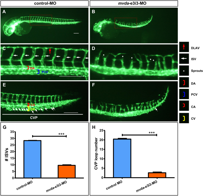
Morpholino knockdown of mvda impairs the trunk angiogenesis and CVP formation in zebrafish. A–F Representative fluorescent images of Tg(fli1a:EGFP)y1 embryos at 2-dpf, with the vascular structures visualized by eGFP fluorescence. The boxed regions of A and B are shown at a higher magnification in C and D, respectively. C ISVs and DLAV showed regular development in the embryos injected with control-MO. D Compared with the control group, embryos injected with mvda-e3i3-MO presented a lower number of incomplete ISVs and ectopic sprouts (asterisks) of DA. E In control embryos, CVP was formed honeycomb-like structures at the tail around 2-dpf (white arrows). F In contrast, mvda knockdown resulted in specific defects in CVP formation. G Quantification of the number of complete ISVs shows a significant decrease in mvda morphants. H Quantification of loop formation at CVP shows a 7.8-fold decrease in mvda morphants. Columns, mean; bars, SEM (n = 10; unpaired student’s t-test) ***P < 0.0001. ISV intersegmental vessel, DLAV dorsal longitudinal anastomotic vessels, DA dorsal aorta, CVP caudal vein plexus, PCV posterior cardinal vein, CA caudal artery, CV caudal vein, dpf days post fertilization. Scale bars = 100 µm
This image is the copyrighted work of the attributed author or publisher, and
ZFIN has permission only to display this image to its users.
Additional permissions should be obtained from the applicable author or publisher of the image.
Full text @ Biol. Res.
Your Input Welcome
Thank you for submitting comments. Your input has been emailed to ZFIN curators who may contact you if
additional information is required.
Oops. Something went wrong. Please try again later.