FIGURE

Fig. 3

ID
ZDB-FIG-210606-14
Publication
Wong et al., 2021 - Mvda is required for zebrafish early development
Other Figures
All Figure Page
Back to All Figure Page
Fig. 3

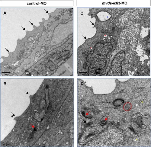
TEM of 4-dpf larvae injected with control or mvda morpholinos. A, B Keratinocytes of the control group revealed regular arranged spicules (black arrows) and cytoplasmic keratin filaments. C, D Keratinocytes of the mvda-e3i3-MO-injected zebrafish revealed disorganized spicules (black arrows), disordered cytoplasmic keratin filaments, distended rough endoplasmic reticulum (yellow asterisks), swollen mitochondria (red circle), autophagosomes and mitophagy (red arrowheads), vesicles (red asterisks), and rupture of cell–cell contacts (blue asterisk). Open arrowheads point to the basement membrane; e, epidermis; d, dermis; cs, collagenous stroma; dpf, days post fertilization. Scale bars = 1 µm (A, C) and 0.5 µm (B, D)

Expression Data

Expression Detail
Antibody Labeling
Phenotype Data
Fish:
Knockdown Reagent:
Observed In:
Stage: Day 4

Phenotype Detail
Acknowledgments
This image is the copyrighted work of the attributed author or publisher, and ZFIN has permission only to display this image to its users. Additional permissions should be obtained from the applicable author or publisher of the image. Full text @ Biol. Res.