IMAGE

Fig. 5

ID
ZDB-IMAGE-210606-21
Source
Figures for Wong et al., 2021
Image
Figure Caption

Fig. 5

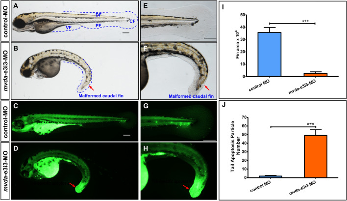
mvda deficiency induces fin-reduction phenotypes and tail-specific apoptosis. A normal fins, control-MO; B fin-reduction phenotypes after mvda-e3i3-MO injected; EF caudal fin is shown at higher magnification. Dashed lines indicate the morphology of fins. Control-MO-injected embryos and embryos injected with mvda-e3i3-MO were stained with acridine orange at 4-dpf. Apoptotic cells are visible as bright green spots, and less bright homogenous green or black staining is unspecific background staining. C, G Control-MO-injected zebrafish exhibited few or no apoptotic cells in whole organism. D, H In contrast, significantly increased staining was observed throughout the tail in zebrafish injected with mvda-e3i3-MO (red arrows). I Quantification of area at fin shows a 13.9-fold decrease in mvda morphants. Error bars, s.e.m.; ***P < 0.0001(n = 10; ANOVA). J Quantification of apoptosis particle number at tail shows a 25.8-fold increase in mvda morphants. Error bars, s.e.m.; ***P < 0.0001(n = 10; Student’s t test); A–H: lateral view, anterior, left. CF caudal fin, DF dorsal fin, PF pelvic fin, VF ventral fin, dpf days post fertilization. Scale bars = 100 µm

Figure Data
Acknowledgments
This image is the copyrighted work of the attributed author or publisher, and ZFIN has permission only to display this image to its users. Additional permissions should be obtained from the applicable author or publisher of the image. Full text @ Biol. Res.