
- Title
-
Anti-Inflammatory Effects of Antarctic Lichen Umbilicaria antarctica Methanol Extract in Lipopolysaccharide-Stimulated RAW 264.7 Macrophage Cells and Zebrafish Model
- Authors
- Hong, J.M., Kim, J.E., Min, S.K., Kim, K.H., Han, S.J., Yim, J.H., Park, H., Kim, J.H., Kim, I.C.
- Source
- Full text @ Biomed Res. Int.
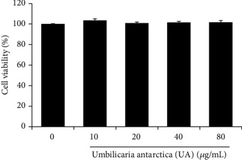
Effect of the methanolic extract of U. antarctica (UA) on the proliferation of RAW 264.7 cells. Cells were treated with various concentrations of UA extract for 24 h, and cell viability was measured by MTT assay. Three independent experiments were performed, and the data are presented as the . |
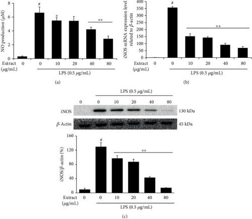
Inhibitory effects of UA extract on nitric oxide (NO) production and inducible-nitric oxide synthase (iNOS) expression in LPS-stimulated RAW 264.7 cells. (a) Cells were pretreated with the indicated concentrations of UA extract for 1 h prior to incubation with LPS (0.5 μg/mL) for 24 h. The nitrite levels in the culture medium were measured by the Griess reaction. Three independent experiments were performed, and the data are presented as the mean ± SEM. *P<0.05 and **P<0.01 compared to the cells treated with LPS alone. (b) Cells were pretreated with UA extract for 1 h and then stimulated with LPS for 6 h. β-Actin expression was used as an internal control for quantitative RT-PCR. (c) Cells were pretreated with UA extract for 1 h and then induced with LPS for 24 h. Ten cell lysates were prepared, and the iNOS protein levels were analyzed on western blots. Expression of β-actin was used as an internal control for western blot analysis. |
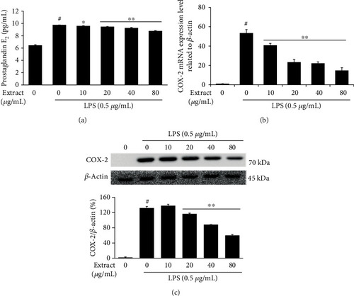
Inhibitory effects of UA extract on prostaglandin E2 (PGE2) production and cyclooxygenase-2 (COX-2) expression in LPS-stimulated RAW 264.7 cells. (a) Cells were treated with the indicated concentrations of UA extract for 1 h prior to incubation with LPS (0.5 μg/mL) for 24 h. The PGE2 levels in the culture medium were measured using enzyme-linked immunosorbent assay (ELISA). Three independent experiments were performed, and the data are presented as the mean ± SEM. *P<0.05 and **P<0.01 compared to the cells treated with LPS alone. (b) Cells were treated with UA extract for 1 h and then induced with LPS for 6 h. β-Actin expression was used as an internal control for quantitative RT-PCR analysis. (c) Cells were treated with UA extract for 1 h and then induced with LPS for 24 h. Ten cell lysates were prepared, and the COX-2 protein expression levels were analyzed by western blotting. β-Actin expression was used as an internal control for western blots. |
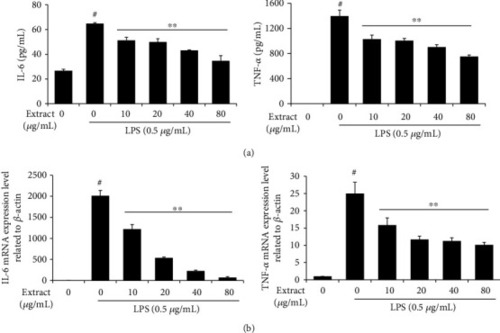
Effects of UA extract on the transcription and translation of interleukin- (IL-) 6 and TNF-α in LPS-induced RAW 264.7 cells. (a) Cells were treated with the indicated concentrations of UA extract for 1 h prior to incubation with LPS (0.5 μg/mL) for 24 h. The level of IL-6 and TNF-α in the supernatant was determined by enzyme-linked immunosorbent assay (ELISA). (b) Cells were treated with UA extract for 1 h and then induced with LPS for 6 h. β-Actin expression was used as an internal control for quantitative RT-PCR analysis. Three independent experiments were performed, and the data are presented as the mean ± SEM. *P<0.05 and **P<0.01 compared to the cells treated with LPS alone. |
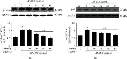
Effect of UA extract on the regulation of the nuclear factor- (NF-) κB pathway. Cells were treated with the indicated concentrations of UA extract for 1 h, followed by induction with LPS (0.5 μg/mL) for 30 min. Equal amounts of protein were analyzed using specific antibodies against p-IκB and p65. Levels of p-IκB and p65 protein in (a) cytosolic and (b) nuclear fractions measured by western blotting. Three independent experiments were performed, and the data are presented as the mean ± SEM. *P<0.05 and **P<0.01 compared to the cells treated with LPS alone. Expressions of GAPDH and PCNA were used as internal controls for western blot analysis. |
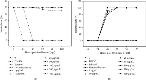
Effect of UA extract on development in zebrafish embryos for 120 hpf. Survival and hatching rates were monitored in zebrafish embryos after exposure at epiboly stage to various concentrations (1, 10, 25, 50, 100, 200, and 400 μg/mL) of UA extract for 5 days. Experiments were performed in three replicates for each concentration. |
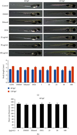
Effects of UA extract on embryonic development of zebrafish. (a) After treatment with various concentrations (0, 25, 50, and 100 μg/mL) of UA extract, the phenotype and body length of zebrafish larvae in 48 hpf and 120 hpf were studied. (b) The effect of UA extract on the heartbeat rate at 48 hpf of zebrafish embryos. |
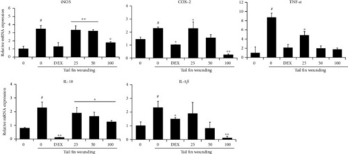
Effect of UA extract on inflammatory gene expression in zebrafish larvae. Tail fin from 3 dpf zebrafish larvae was wounded, and mRNA levels of iNOS, COX-2, TNF-α, IL-10, and IL-1β were determined by quantitative real-time PCR. Gene expression was normalized against that of β-actin. Data represent the average of the three replicates, and P values were calculated using t-test. *P<0.05 and **P<0.01. |
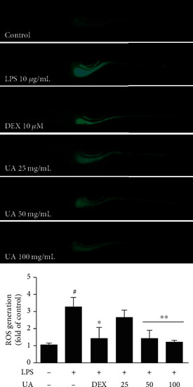
Effect of UA extract on LPS-induced ROS generation in zebrafish larvae. (a) Zebrafish larvae were treated with 25, 50, and 100 μg/mL UA extract and 10 μg/mL LPS for 24 h. The levels of ROS were observed under a fluorescence microscope after staining with DCF-DA. (b) The fluorescence intensities in individual zebrafish larvae were quantified. Data represent the average of the three replicates, and P values were calculated using t-test. *P<0.05 and **P<0.01. |