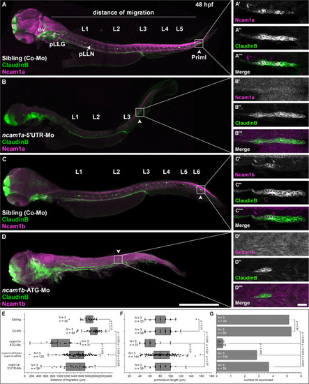
Expression pattern and function of Ncam1 during development of the zebrafish lateral line system. (A–D) Lateral views of Tg(ClaudinB::lynGFP) (green) embryos (48 hpf) immunostained for Ncam1a or Ncam1b (magenta). (A,C) Control-Morpholino (Co-Mo) injected sibling; all structures (excluding interneuromast cells) of the lateral line system express Ncam1a and Ncam1b. (A′-A,C′-C) Ncam1a is expressed throughout the whole primordium (arrowhead), whereas Ncam1b is found only in the Trailing-Zone. (B)ncam1a-morphants show only weak migration defects and (B′-B) primordium length is not affected. (D) Morpholino-knockdown of ncam1b induces migration defects as well as a reduction in the number of deposited (pro)neuromasts. (D′-D)ncam1b-knockdown also results in a reduced primordium size. (E,F,G) Quantification of migration distance, primordium length and number of neuromasts in control and morpholino-injected embryos. Rescue mRNA was co-injected with the morpholino as indicated. Error bars represent standard deviation. Scale bars (A,D) 200 μm, (D) 20 μm. Abbreviations: Co-Mo, Control-Morpholino; L1-6, lateral (pro)neuromast; pLLG, lateral line ganglion; pLLN, lateral line nerve; ov, otic vesicle; PrimI, primary posterior primordium.

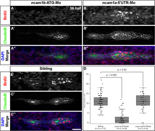
Ncam1b is required for cell proliferation within the primordium. Siblings or morpholino-injected Tg(ClaudinB::lynGFP) embryos were treated with BrdU at 36 hpf for 20 min, fixed and immunostained for BrdU (red), GFP (green) and DAPI (blue) as indicated. (A,A″) BrdU incorporation is almost absent in ncam1b-morphants, but obvious in (B,B″) knockdowns of ncam1a and (C,C″) uninjected siblings. (D) Quantification of BrdU-labeled cells within primordia. Scale bar 20 μm.

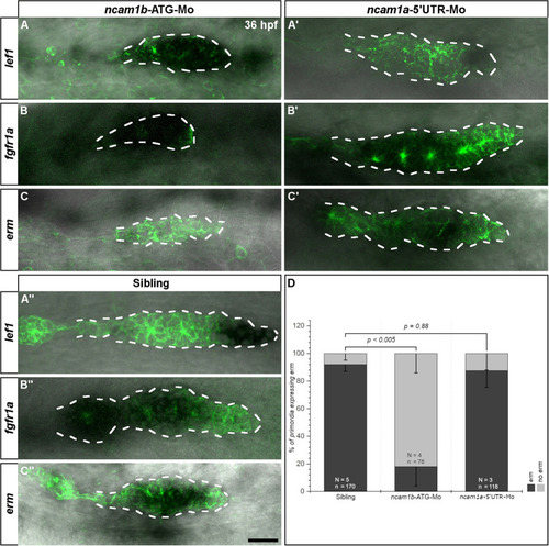
Ncam1b affects the expression of the Fgfr1a-target gene erm. Tg(ClaudinB::lynGFP) embryos were immunostained labeled for GFP (green). In situ hybridizations of probes detecting lef1 (Leading-Zone), fgfr1a, or erm (both in the Trailing-Zone) were visualized by NBT/BCIP staining. Primordia outlines are surrounded with a dotted line for better visibility. (A–C) Neither expression of lef1 nor of fgfr1a are affected in ncam1b-morphants, whereas erm expression is markedly reduced. (A′,C′) Neither ncam1a-morphants nor (A″,C″) siblings show alterations for lef1, fgfr1a, or erm. (D) Reduced erm expression after knockdown of ncam1b. Error bars show standard deviation. X2-test with ns = p0.05. Scale bar 20 μm.

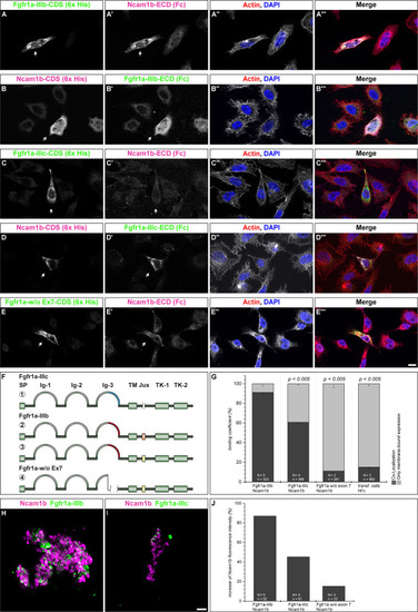
Ncam1b interactions with different isoforms of Fgfr1a. CHO-K1 cells were transfected with complete coding sequences (CDS) of one of three Fgfr1a-isoforms (green; IIIb, IIIIc, or w/o Ex7) or of Ncam1b (magenta). Transfected cells were incubated for 1 hr with either soluble Ncam1b or one of the two Fgfr1a-isoforms, fixed and immunostained for His-, Fc-Tag, Actin (red), and DAPI (blue). (A,B) Ncam1b interacts strongly with Fgfr1a-IIIb irrespectively of which of the proteins is membrane-bound. (C,D) Binding of Ncam1b to Fgfr1a-IIIc is markedly weaker. (E,E) No interaction occurs if Fgfr1a lacks exon 7. (F) Scheme of isolated and cloned Fgfr1a splice variants: ① Fgfr1a-IIIc does not contain any exon 9, ② one splice variant of Fgfr1a-IIIb contains a juxtamembrane region of valine and threonine (orange), ③ another isoform of Fgfr1a-IIIb encodes two serine residues (yellow) within the juxtamembrane domain, ④ Fgfr1a-IIIa, an artificial variant of Fgfr1a, lacks the C-terminal part of Ig-3, which is encoded by exon 7. (G) Quantification of the binding of Ncam1b to splice variants of Fgfr1a. Binding coefficient represents the percentage of cells expressing the membrane-bound binding partner that were co-labeled by antibodies against the soluble binding partner. Error bars represent standard deviations (χ2-test). (H,I) Bead aggregation assays reveal a strong homophilic binding of Ncam1b. Ncam1b-coated beads interact with Fgfr1a-IIIb-coated beads and to a lower extend with Fgfr1a-IIIc. (J) Measuring the fluorescence intensity of soluble Ncam1b-Fc shows a strong binding to membrane-bound Fgfr1a-IIIb and a weaker to Fgfr1a-IIIc or Fgfr1a without exon 7. Scale bar in (E) represents 20 μm and in (I) 10 μm. Abbreviations: CDS, coding sequence; ECD, extracellular domain; lg, Immunoglobulin domain; Jux, juxtamembrane domain; SP, Signal peptide; TK, Tyrosine Kinase domain.

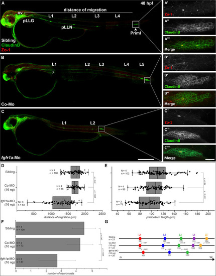
Fgfr1a is important for posterior lateral line development. Lateral views of 48 hpf Tg(ClaudinB::lynGFP) (green) embryos. (A,B) In siblings and control-morpholino injected embryos Priml has reached the tip of the tail and deposited 5 neuromasts. (C)fgfr1a-knockdown embryos show a delayed migration of primordia and a reduced number of neuromasts. (A′-C′) Apical constriction, visualized by staining of Zo-1, occurs under all conditions. Quantification of (D) migration distance, (E) primordium length, (F) number and (G) spacing of neuromasts, respectively. Error bars in bar chart represent standard deviation. Scale bars (A,B) 200 μm, (A′-C″) 20 μm. Abbreviations: Co-Mo, Control-Morpholino; L1-6, lateral (pro)neuromast; pLLGg, lateral line ganglion; pLLN, lateral line nerve; ov, otic vesicle; PrimI, primary posterior primordium.

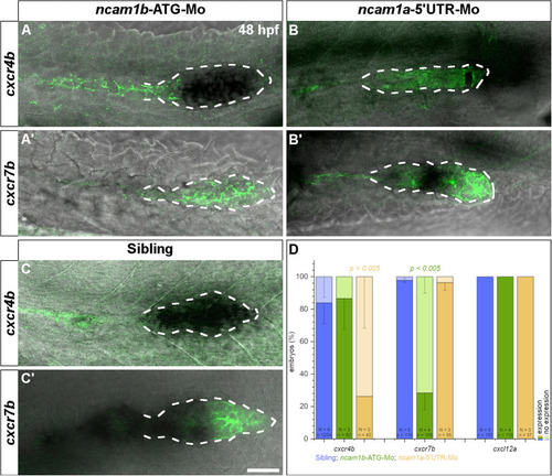
Expression of chemokine receptor cxcr7b is affected by knockdown of ncam1b. Tg(ClaudinB::lynGFP) embryos (48 hpf) were immunostained for GFP (green). In situ hybridizations of probes detecting cxcr4b (complete PrimI) and cxcr7b (Trailing-Zone), were visualized by NBT/BCIP staining. Primordia outlines were surrounded with a dotted line for better visibility. (A,A′) Expression of cxcr4b is not affected in ncam1b-morphants, whereas expression of cxcr7b is markedly reduced. (B,B′) In ncam1a-morphants cxcr7b is present, whereas cxcr4b expression is reduced. (C,C′) In siblings, cxcr7b is robustly expressed in the Trailing-Zone. (D) Drastically reduced cxcr7b expression after knockdown of ncam1b. Error bars represent standard deviation. X2-test with ns = p0.05. Scale bar 20 μm.

The function of Ncam1b during posterior lateral line development. (A) Expression patterns of Ncam1b, Fgfr1a, Wnt, Erm and Lef1 in the pLLS primordium. (B) Interaction of Ncam1b with Fgfr1a results in activation of the transcription factor Erm, which is involved in cell proliferation in the Trailing-Zone of PrimI. The Ncam1b/Fgfr1a interaction also initiates the expression of the chemokine receptor Cxcr7b, which is necessary for directional migration of the PrimI and for proneuromast deposition. Wnt signaling controls proliferation in the Leading-Zone via Lef1. In addition, it triggers Fgf signaling in the Trailing-Zone, which initiates apical constriction during formation of proneuromasts. Proneuromasts are stabilized by homophilic Ncam1b binding in trans. (Data related ① to Lecaudey et al. (2008) ② Aman et al. (2011) ③ Aman and Piotrowski (2008).

Acknowledgments
This image is the copyrighted work of the attributed author or publisher, and ZFIN has permission only to display this image to its users. Additional permissions should be obtained from the applicable author or publisher of the image. Full text @ Front Cell Dev Biol