
- Title
-
Synchronized tissue-scale vasculogenesis and ubiquitous lateral sprouting underlie the unique architecture of the choriocapillaris
- Authors
- Ali, Z., Cui, D., Yang, Y., Tracey-White, D., Vazquez-Rodriguez, G., Moosajee, M., Ju, R., Li, X., Cao, Y., Jensen, L.D.
- Source
- Full text @ Dev. Biol.

Early development of the choriocapillaris in zebrafish. A: Time lapse confocal micrographs of the developing choriocapillaris (vessels shown in green) of the outer eye field in Fli1a:EGFP embryos at 18–23 h post fertilization (hpf, top row) and 24–120 hpf (lower row). Boxed regions are shown in magnified images below. Yellow arrows point to the indicated vessel structures, yellow arrowheads points to the avascular area or interstitial foci while white arrowheads points to endothelial cells or vessels. The dotted line indicates the eye field. Size bars indicates 50 μm in each image. CrDI: Cranial division of internal carotid artery, HA: Hyaloid artery, OA: Optic artery, PMBC: Primordial midbrain channel. DRV: Dorsal radial vessel. NRV: Nasal radial vessel. B: Quantifications of the number of the endothelial cells in the experiment shown in A. n = 10–15 embryos, **: p < 0.01 ***: p < 0.001. C: Quantification of the number of interstitial foci in the experiment shown in A. n = 10–15 embryos, ***:p < 0.001. D: Quantification of the vascular density of choriocapillaris in the experiment shown in A. n = 10–15 embryos, ***:p < 0.001. E: Imaris 3D rendering images of the choriocapillaris showing the lumen of CCs of complete intussusceptive pillars (red arrow) and incomplete pillars (white arrows) from 72, 96 and 120 hpf. Size bar indicates 10 μm. hpf: hours post fertilization. F: Scheme illustrating the development of choriocapillaris from 24 hpf to 120 hpf. Development of the choriocapillaris begins with the invasion of the endothelial cells via the PMBC and CrDi at 18–24 hpf. At 36 hpf the endothelial cells forms aggregates (blood islands) interconnected by thin processes (EC-EC connections), leading to formation of a primitive, non-lumenized vasculature at 48 hpf. The choriocapillaris become perfused at 72 hpf but remains actively expanding until maturation occurs at 120 hpf. |

Maturation of choriocapillaris leads to increased perfusion and reduced leakage. A: Confocal micrographs of the posterior eye of Kdrl:EGFP; Gata1:DsRed double transgenic embryos (endothelium shown in green and erythroblasts shown in red) at 48–120 hpf. Boxed regions are shown in magnified images below. White arrowheads points to erythrocytes. Size bar indicates 50 μm hpf, hours post fertilization. D: Quantification of the number of erythrocytes in the choriocapillaris between 48 and 120 hpf from the experiment shown in A. n = 10–15 embryos, ***:p < 0.001. C: Confocal micrographs of Kdrl:EGFP embryos (endothelium shown in green) at different time points between 48 and 120 hpf, 1 h after injection with rhodamine-conjugated 70 kDa dextran via the common cardinal vein (dextran (Dex) shown in red). Boxed regions are shown in magnified images below. Yellow arrowheads points to dextran leaked into the avascular area while white arrowheads points to dextran contained within the vascular lumen. Size bar indicates 50 μm. D: Quantification of perfusion in the choriocapillaris between 48 and 120 hpf. n = 10–15 embryos, ***:p < 0.001. E: Quantification of the dextran leakage between 48 and 120hpf. n = 10–15 embryos, ***:p < 0.001. |

Ultrastructure analysis of the choriocapillaris over development. Transmission electron micrographs (TEM) of 3–5 days post fertilization (dpf) zebrafish outer retina and choroid. Boxed regions are shown in the magnified images below. Bm: Bruch's membrane, F. Fenestration, L. Lumen. Size bar indicates 5 μm. |

Appearance of Rete Mirabile in the zebrafish during development. A: Bright field micrographs from cross sections of 4 weeks and adult zebrafish eyes stained with toluidine blue. The boxed regions are shown in the magnified images below. Size bars indicate 250 μm in each image. CCs. Choriocapillaries, RM. Rete Mirabile. B: Confocal micrographs of the choriocapillaris and Rete Mirabile from Fli1a:EGFP transgenic zebrafish (endothelium shown in green) at 2, 3, 4 weeks and adult stages. Yellow arrows indicate avascular area or interstitial foci and white arrows indicates vascular area. Size bars indicate 20 μm in each image. |
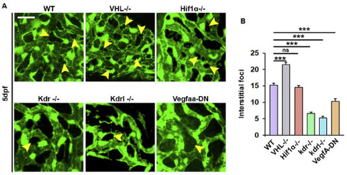
Development of choriocapillaris is dependent on VEGF-VEGFR2 in zebrafish. A: Confocal micrographs of the choriocapillaris of 5 days post fertilization (dpf) embryos from wildtype (WT), VHL mutant (VHL-/-), hif1aa-/-;hif1ab-/- (HIF1a-/-), Vegfr2b-/- (kdr -/-), Vegfr2a-/- (kdrl -/-) or fli1a:EGFP; Hsp70:Vegfaa-DN zebrafish incubated for 1 h at 37 °C daily from 1 to 5 dpf (Vegfaa-DN) crossed onto the Fli1a:EGFP background (endothelium shown in green). Yellow arrowheads indicate interstitial foci. Size bars indicates 20 μm in each image. B: Quantification of the number of interstitial foci in the experiment shown in A. n = 10–15 embryos, ***:p < 0.001. ns: non-significant. |
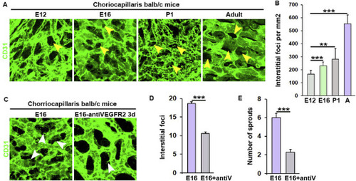
Development of the choriocapillaris in mice is dependent of VEGFR2 signaling. A: Confocal micrographs of the choriocapillaris (CCs) from embryonic day (E)12, 16, postnatal day (P)1 and adult BALB/c mice stained with anti-CD31 (endothelium shown in green). Yellow arrowheads indicate interstitial foci. B. Quantification of the number of interstitial foci in the experiment shown in A. n = 10–15 areas of interest from four embryos collected from two litters, ***:p < 0.001, **:p < 0.005. A: Adult. C. Confocal micrographs of the choriocapillaris from E16 BALB/c mice treated with antiVEGFR2 neutralizing antibody or control IgG antibody for 3 days before staining with anti-CD31 antibody and visualization on the right side. D: Quantification of the number of interstitial foci in the experiment shown in C. n = 10–15 areas of interest from four embryos collected from two litters, ***:p < 0.001. antiV: Anti-VEGFR2-treated. E: Quantification of the number of sprouts in the experiment shown in C. n = 10–15 areas of interest from four embryos collected from two litters, ***:p < 0.001. |
Reprinted from Developmental Biology, 457(2), Ali, Z., Cui, D., Yang, Y., Tracey-White, D., Vazquez-Rodriguez, G., Moosajee, M., Ju, R., Li, X., Cao, Y., Jensen, L.D., Synchronized tissue-scale vasculogenesis and ubiquitous lateral sprouting underlie the unique architecture of the choriocapillaris, 206-214, Copyright (2019) with permission from Elsevier. Full text @ Dev. Biol.