
Image
|
Figure Caption
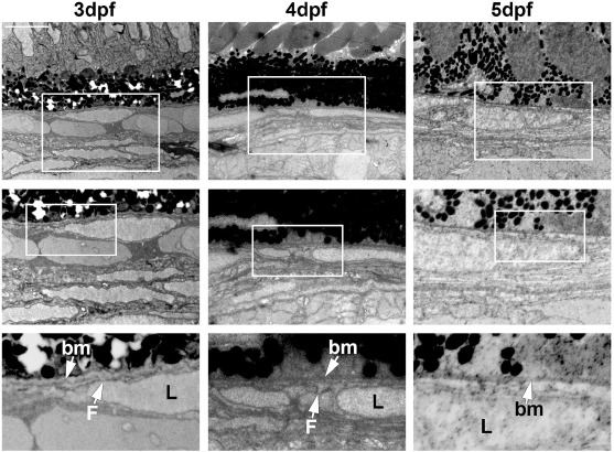
Fig. 3 Ultrastructure analysis of the choriocapillaris over development. Transmission electron micrographs (TEM) of 3–5 days post fertilization (dpf) zebrafish outer retina and choroid. Boxed regions are shown in the magnified images below. Bm: Bruch's membrane, F. Fenestration, L. Lumen. Size bar indicates 5 μm.
Acknowledgments
This image is the copyrighted work of the attributed author or publisher, and
ZFIN has permission only to display this image to its users.
Additional permissions should be obtained from the applicable author or publisher of the image.
Reprinted from Developmental Biology, 457(2), Ali, Z., Cui, D., Yang, Y., Tracey-White, D., Vazquez-Rodriguez, G., Moosajee, M., Ju, R., Li, X., Cao, Y., Jensen, L.D., Synchronized tissue-scale vasculogenesis and ubiquitous lateral sprouting underlie the unique architecture of the choriocapillaris, 206-214, Copyright (2019) with permission from Elsevier. Full text @ Dev. Biol.