
|
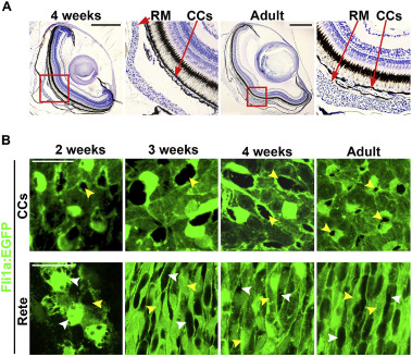
Fig. 4 Appearance of Rete Mirabile in the zebrafish during development. A: Bright field micrographs from cross sections of 4 weeks and adult zebrafish eyes stained with toluidine blue. The boxed regions are shown in the magnified images below. Size bars indicate 250 μm in each image. CCs. Choriocapillaries, RM. Rete Mirabile. B: Confocal micrographs of the choriocapillaris and Rete Mirabile from Fli1a:EGFP transgenic zebrafish (endothelium shown in green) at 2, 3, 4 weeks and adult stages. Yellow arrows indicate avascular area or interstitial foci and white arrows indicates vascular area. Size bars indicate 20 μm in each image.
Reprinted from Developmental Biology, 457(2), Ali, Z., Cui, D., Yang, Y., Tracey-White, D., Vazquez-Rodriguez, G., Moosajee, M., Ju, R., Li, X., Cao, Y., Jensen, L.D., Synchronized tissue-scale vasculogenesis and ubiquitous lateral sprouting underlie the unique architecture of the choriocapillaris, 206-214, Copyright (2019) with permission from Elsevier. Full text @ Dev. Biol.