- Title
-
The Serotonergic System Tracks the Outcomes of Actions to Mediate Short-Term Motor Learning
- Authors
- Kawashima, T., Zwart, M.F., Yang, C.T., Mensh, B.D., Ahrens, M.B.
- Source
- Full text @ Cell
|
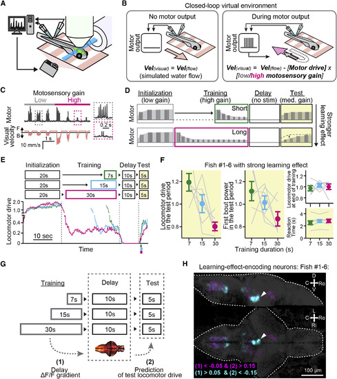
Whole-Brain Activity Mapping of Learning-Effect-Encoding Neurons (A) Experimental setup. Signals from motor neuron axons (fictive swimming) trigger real-time visual feedback. A light-sheet microscope images the brain. (B) Virtual environment. When the fish does not fictively swim (left), a visual scene moves forward slowly to elicit swimming. Fictive swim signals (right, inset) move the visual scene backward to mimic the effect of forward swimming. The speed of visual feedback is controlled by the motosensory gain. For the same swim bout, high gain yields more visual flow (magenta arrow) than low gain (gray arrow). (C) Example swim trace and visual motion during a transition from low to high motosensory gain. “Motor” signal is the power in a sliding window of the tail voltage signal. Insets show zoom-ins of boxed regions. (D) The short-term motor learning paradigm consists of repetitions of the initialization, training, delay, and test periods. No time passes during arrows between periods. Schematized electrophysiology traces represent the strength of fictive swim bouts. The learning effect is the dependence of locomotor drive in the test period on the duration of training. (E) Representative fish behavior. Longer high-motosensory-gain training (magenta) more strongly attenuates locomotor drive in the test period. Colored lines below the x axis of the test period, mean ± SD of reaction times. (F) Behavior across six fish with strong learning effect. Left: locomotor drive in the test period (average integrated swim power in the test period normalized by the average across three training conditions) is more attenuated after longer training. Right: locomotor drive at the end of training (top, last 5 s, normalized across conditions) and reaction time to the test stimulus (bottom) are similar across training durations. Gray lines, data from individual fish. Error bars, SEM across fish. Plots and statistics for all fish are in Figures S1B and S1C. (G) Whole-brain analysis. Encoding of the learning effect was quantified by (1) dependence of a neuron’s average ΔF/F activity signal in the delay period on the preceding training duration, and (2) degree to which a neuron’s ΔF/F in the delay period predicts the locomotor drive in the subsequent test period (test locomotor drive) trial-by-trial. (H) Functional brain map of neurons with parameters (1) and (2) exceeding thresholds, overlay from six fish with strong learning effect. A density of neurons occurs in the DRN (white arrow). Thresholds for extracting neurons are indicated at the bottom left (cyan for positive correlation to the learning effect; magenta for negative). See also Figures S2 and S3 and Movie S1. |
|
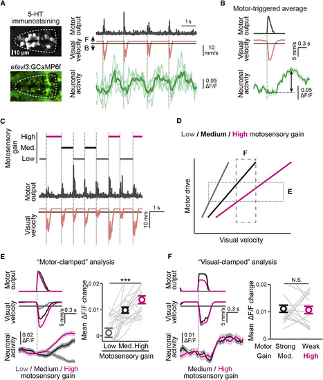
DRN Neurons Encode Sensory Feedback of Motor Action (A) High-speed imaging of DRN neurons. Left: 5-HT immunostaining and GCaMP6f. Right: fictive swim signal, visual motion and ΔF/F activity of four group 1 DRN neurons (thick line is average ΔF/F). (B) Example DRN neuron activity. Top: average locomotor drive across swim events aligned to bout onsets (dotted gray line). Middle: average visual velocity. Bottom: average ΔF/F of a group 1 DRN neuron. Black arrow: response magnitude used for analysis. Shadows, SEM across swim events. (C) Stochastic gain paradigm. In a closed-loop environment, at every bout (gray, middle), motosensory gain is set randomly to low (dark gray), medium (black) or high (magenta). (D) Analysis for (E) and (F). In motor-clamped analysis (E), visual coding is quantified by the dependence of the response on visual velocity within a restricted range of locomotor drive (data within horizontal bar). In visual-clamped analysis (F), motor coding is quantified by the dependence on locomotor drive within a restricted range of visual velocity (vertical bar). (E) Motor-clamped analysis of DRN neurons’ visual responses during the stochastic gain paradigm (see D). Left, average swim trace (top), visual motion (middle) and ΔF/F of a group 1 DRN neuron (bottom) under different motosensory gains. Right, dependence of group 1 DRN neurons’ responses under different motosensory gains. ∗∗∗p = 4.7 × 10⁻⁹ for effect of motosensory gain by linear mixed-effects model (LME), 21 fish. Error bars, SEM across fish. Gray lines, data from individual fish. (F) Visual-clamped analysis of motor-related responses of group 1 DRN neurons (see D). Left, example neuron with similar responses to events with different swim power but similar visual motion. Right, responses of DRN neurons across fish. Average response strength of group 1 neurons does not depend on locomotor drive. Error bars, SEM, 15 fish. Gray lines, data from individual fish. See also Figure S4. |
|
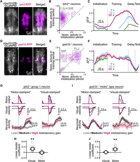
Cell-Type-Specific DRN Dynamics (A–C) Dynamics of serotonergic DRN neurons during the motor learning paradigm. (A) Double labeling of DRN neurons with H2B-GCaMP6f (gray) and serotonergic DRN neurons with RFP (magenta). (B) Activity in the initialization and training periods of 420 serotonergic neurons across seven fish, same analysis as in Figure 4A. Activity is higher during training for most neurons. (C) Dynamics of serotonergic neurons during formation and retention of the learning effect of neurons in box C in (B), averaged over 11 trials and across 94 tph2+ DRN neurons in one representative fish. Shadows, SEM across neurons. (D–F) Dynamics of GABAergic DRN neurons during the motor learning paradigm. (D) Double labeling of DRN neurons with H2B-GCaMP6f (gray) and GABAergic neurons with RFP (magenta). (E) Activity in the initialization and training periods of 356 neurons from nine fish. (F) Dynamics of GABAergic neurons in box F in (E), averaged over 11 trials and across 47 GAD1B+ neurons for one representative fish, showing activation when fish swims (initialization, training, test) and rapid decay when it does not (delay). Shadows, SEM across neurons. (G–J) Neural activity imaged at 30 Hz during the stochastic gain paradigm of Figure 2C. Sources of population data are indicated by boxes in (B) and (E). (G) Responses of an example group 1 serotonergic neuron represented by (left) dependence on visual stimulus velocity by motor-clamped analysis as in Figure 2E and (right) dependence on locomotor drive by visual-clamped analysis as in Figure 2F. (H) Linear model fit to predict response amplitude from swim power and visual input confirms that average responses of serotonergic group 1 neurons are primarily tuned to visual stimulus velocity and less to locomotor drive as in Figures 2E and 2F and consistent with the response profile in (G). ∗∗p = 0.0014 by one-sample t test, 11 fish. Error bars: SEM across fish. (I) Responses of an example GABAergic motor-type neuron by (left) dependence on visual velocity by motor-clamped analysis as in Figure 2E and (right) dependence on locomotor drive by visual-clamped analysis as in Figure 2F. (J) Fitting a linear model as in (H) confirms that average responses of GABAergic motor-type neurons are primarily tuned to locomotor drive and less to visual stimulus velocity, consistent with response profile in (I). ∗∗p = 0.0052 by one-sample t test, eight fish. Error bars: SEM across fish. See also Figure S5. |
|
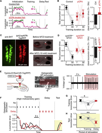
Serotonin Transmission and the DRN Are Causally Related to Learning (A and B) Effects of pCPA serotonin release block on the learning effect. (A) Behavioral trace of control (top) and pCPA-treated (bottom) fish during the motor learning paradigm. (B) pCPA treatment leads to a loss of the learning effect. Left: test locomotor drive (integrated locomotor drive in the test period, normalized by the average across training conditions) is attenuated by longer training in the control group (black), but not in the pCPA-treated group (red). Faint lines, data from individual fish. Right: differences between test locomotor drive after short (7 s) or long (30) training durations. ∗p = 0.017 by Wilcoxon rank-sum test between ten control fish and ten pCPA-treated fish. Error bars, SEM across fish. (C and D) Effect of chemical-genetic ablation of tph2+ serotonergic neurons on motor learning. (C) Left: 5-HT immunostaining (green) and nitroreductase (epNTR) expression in a Tg(tph2:epNTR-RFP) fish (red) in the DRN. Right: epNTR-TagRFP expression in the DRN (arrowhead) taken before (top, 4 dpf) and after (bottom, 6 dpf) metronidazole (MTZ) treatment for the same fish, fluorescence overlaid on bright field images. Most DRN neurons disappear. (D) Chemical genetic ablation leads to loss of the learning effect. Left: locomotor drive in the test period is attenuated by longer training in epNTR− siblings (black), but not in epNTR+ group (red) after MTZ treatment. Faint lines, data from individual fish. Right: differences between test locomotor drive after short (7 s) and long (30 s) training. ∗p = 0.046 by Wilcoxon rank-sum test between eight epNTR− fish and nine epNTR+ fish. Error bars, SEM across fish. (E) Two-photon optogenetic activation of DRN neurons. Left: DRN neurons are specifically activated by laser scanning over a plane within the DRN. Center: location of a DRN neuron recorded by loose patch and electroporated with dye after recording. Red box, scan area. Right: raw recorded trace before and during optogenetic activation (red shadow) showing that the laser elicits extra spikes. (F) Instantaneous effects of DRN activity on locomotor drive tested by stimulation during training (gray vertical lines and behavioral trace). Lasting effects were tested by stimulating in the delay period (red). (G) DRN stimulation suppresses ongoing locomotor drive. Training locomotor drive is the integrated drive in the training period, normalized to the average across three stimulation conditions. p = 0.027 by one-way ANOVA. ∗p = 0.038 by Tukey’s post hoc test. Error bars, SEM, nine fish. Gray dotted lines, data from individual fish. (H) DRN stimulation in the delay period attenuates drive in the subsequent test period. This effect bridges the gap of ∼3 s between the last stimulation pulse and the first swim bout in the test period. p = 0.0055 by one-way ANOVA. ∗∗p = 0.0069 by Tukey’s post hoc test between delay stimulation and no stimulation for same nine fish as (G). Error bars, SEM across fish. Gray dotted lines, data from individual fish. See also Figures S6 and S7. |
|
Anatomy of Serotonergic Neurons in the DRN, Related to Figure 1 (A) Distribution of 5-HT positive neurons in the hindbrain. Left, schematic diagram of hindbrain region imaged with a confocal microscope. Right, maximum intensity projection of 5-HT immunostaining from the side (top) and the top (bottom) of the hindbrain. The side view is a maximum intensity projection of the tissue between the dashed gray lines in the top projection. Serotonergic clusters were classified and labeled above the top panel according to previous literature ( Lillesaar et al., 2009 and Parker et al., 2013). D, dorsal; V, ventral; Ro, rostral; C, caudal; Ri, right; L, left. (B) Left, effect of training duration on behavior in the test period, as described in Figures 1D–1F, in the fish group with weak/no learning effect. Error bars: SEM across fish. Gray lines represent data from individual fish. Right, the overlaid map of learning-effect-encoding neurons in a fish group with weak/no learning effect at the behavioral level, showing far fewer neurons (53 ± 6 and 68 ± 11 neurons per fish for cyan and magenta neurons, respectively) that reach criterion compared to fish with strong learning effect in Figure 1H (206 ± 26 and 220 ± 33 neurons per fish for cyan and magenta neurons, respectively). Neurons are extracted by the same parameters (1) and (2) as explained in the main text and Figures 1G and 1H. D, dorsal; V, ventral; Ro, rostral; C, caudal; Ri, right; L, left. |
|
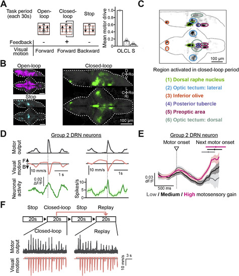
Whole-Brain Map of Neurons that Are Responsive to Closed-Loop Visual Feedback, and Activity Patterns of Group 2 DRN Neurons, Related to Figures 2 and 3 (A) The behavioral task for identifying neurons activated by closed-loop sensory feedback. Left, illustration of the behavioral task. In the ‘open-loop’ period, the visual environment moves forward without any feedback. In the ‘closed-loop’ period, the visual environment moves forward, but when a swim signal is detected it moves backward. In the ‘stop’ period, the visual environment slowly moves backward, during which the fish tend to swim less or cease swimming. Right, normalized locomotor drive in the open-loop (OL), the closed-loop (CL) and the stop (S) period. Locomotor drives of individual fish are normalized to their average locomotor drive in the open-loop period. Error bars represent SEM across 5 fish. (B) Whole-brain maps of neurons that showed highest activity in the open-loop period (magenta, left top), in the closed-loop period (green, right) and in the stop period (cyan, left bottom) morphed and overlaid on a reference brain (gray). n = 5 fish. See the STAR Methods for details of analyses. D, dorsal; V, ventral; Ro, rostral; C, caudal; Ri, right; L, left. (C) Anatomical segmentation of brain regions that showed highest activity in the closed-loop period. Top, the same whole-brain map in (B) inverted for brightness and overlaid with anatomical masks. Bottom, a list of 6 identified anatomical regions. (D) Top, a representative average ΔF/F activity trace (green, bottom left) and a representative electrophysiologically recorded spiking pattern (green, bottom right) from two group 2 DRN neurons recorded separately. Average swim trace is shown in gray (top) and the average visual motion in the virtual reality environment is shown in orange (middle). Gray dotted lines represent the onsets of swim bouts to which the averaged traces are aligned. Shadows represent SEM across averaged swim events. (E) Average ΔF/F activity trace of a representative group 2 neuron in the DRN in response to different motosensory gains during the stochastic gain paradigm. The delayed response is stronger under high motosensory gain (magenta) than under medium (black) or low (dark gray) motosensory gain. The onset time (mean ± SD) of the next swim bout is indicated on the top. For each swim bout, only the activity trace up to the onset of the following bout is included in the average. (F) Behavioral task for desynchronization of swim bouts and visual flow as described in Figure 3B. In the ‘stop’ period of 20 s, the visual motion is stopped. In the ‘closed-loop’ period of 20 s, the visual environment moves forward, but when a swim signal is detected, it moves backward with high motosensory gain. In the ‘replay’ period of 20 s, the same visual motion (orange, bottom) as in the preceding closed-loop period is replayed and the swim events (gray, middle) are desynchronized with visual motion as also shown in Figure 3B. |
|
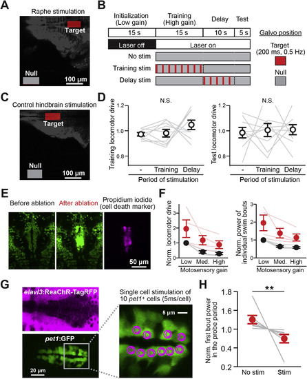
Optogenetic Activation and Laser Ablation of DRN Neurons, Related to Figure 6 (A) ROI settings for DRN stimulation. To stimulate DRN neurons, the target region is scanned, and to remove DRN stimulation, the null region is scanned (as an alternative to using a laser shutter, to avoid auditory stimulation by the rapid opening and closing of the shutter). (B) Detailed stimulation protocol of the experiment presented in Figures 6E–6H. The default laser scan area was set to the ‘Null’ ROI outside the fish. During the period of stimulation, the laser scan area was moved to the ‘Target’ ROI for a short period of time (200 ms, performing four area scans of 50 ms each) once every 2 s. (C) ROI settings for control hindbrain stimulation. (D) Effect of control hindbrain stimulation on the locomotor drive in the training period (left) and the test period (right), showing no significant effects. N.S., p = 0.15 (left) and p = 0.92 (right) by one-way ANOVA across 9 fish. Error bars represent SEM across the same set of 9 fish as in Figures 6G and 6H. Gray lines represent data from individual fish. (E) Two-photon plasma-mediated laser ablation of DRN neurons in Tg(elavl3:H2B-GCaMP6f)jf7 transgenic zebrafish. Dozens of cells in the DRN across multiple Z-planes were ablated; displayed here are images from a single plane. After ablation, the basal intensity of GCaMP fluorescence increased, indicating cell damage. Cell death and its local confinement were further verified post hoc by staining with propidium iodide, a membrane-impermeable dye-specific to dead cells. (F) Locomotor drive (left) and power of individual swim bouts (right) under various levels of motosensory gain before (black) and after (red) DRN cell ablation, showing an increase in locomotor drive following ablation. The fish are still able to perform real-time motor adaptation, which we also observed in the chemical genetic ablation results in Figures 6 and S6. Values were normalized to the average locomotor drive under low motosensory gain before ablation in individual fish. Error bars represent SEM across 4 tested fish. Two-way ANOVA was performed on the effect of ablation and motosensory gain. (Left) p = 0.0045 and (Right) p = 0.0007 between before and after ablation. No interaction in the two-way ANOVA test was detected between the effects of ablation and motosensory gain. Thin red lines and gray lines represent data from individual fish before and after ablation, respectively. (G) Cell-type specific two-photon optogenetic activation of individual serotonergic neurons in the DRN. Double transgenic zebrafish which express ReaChR-TagRFP under the elavl3 promoter (left top, magenta) and GFP in pet1+ serotonergic neurons (left bottom, green) [Tg(elavl3:ReaChR-TagRFP-T; pet1:GFP)] were used in this experiment. 10 pet1+ serotonergic neurons in the DRN were stimulated individually by local spiral scanning, with the scan pathway shown in magenta on the right. These neurons were stimulated 4 times in a 200 ms period (i.e., at 20 Hz within this window), with 5 ms spiral time per cell, timed to be 2 s before the end of the delay period (i.e., about 3 s before the first swim bout in the test period). (H) Effect of stimulation during the delay period on the power of the first bout in the test period. The behavioral paradigm of this experiment is the same as the one in Figure 6F except that we only tested two conditions, control (No stim) and stimulation (Stim) as described in (G). ∗∗p = 0.0078 from one-tailed Wilcoxon signed-rank test across 7 fish for the effect of suppression after stimulation. Gray lines represent data from individual fish. Error bars: SEM across fish. |
Reprinted from Cell, 167, Kawashima, T., Zwart, M.F., Yang, C.T., Mensh, B.D., Ahrens, M.B., The Serotonergic System Tracks the Outcomes of Actions to Mediate Short-Term Motor Learning, 933-946.e20, Copyright (2016) with permission from Elsevier. Full text @ Cell

