
Fig. S7
- ID
- ZDB-IMAGE-161227-17
- Publication
- Kawashima et al., 2016 - The Serotonergic System Tracks the Outcomes of Actions to Mediate Short-Term Motor Learning
- All Figures
- Figures for Kawashima et al., 2016
|
Fig. S7
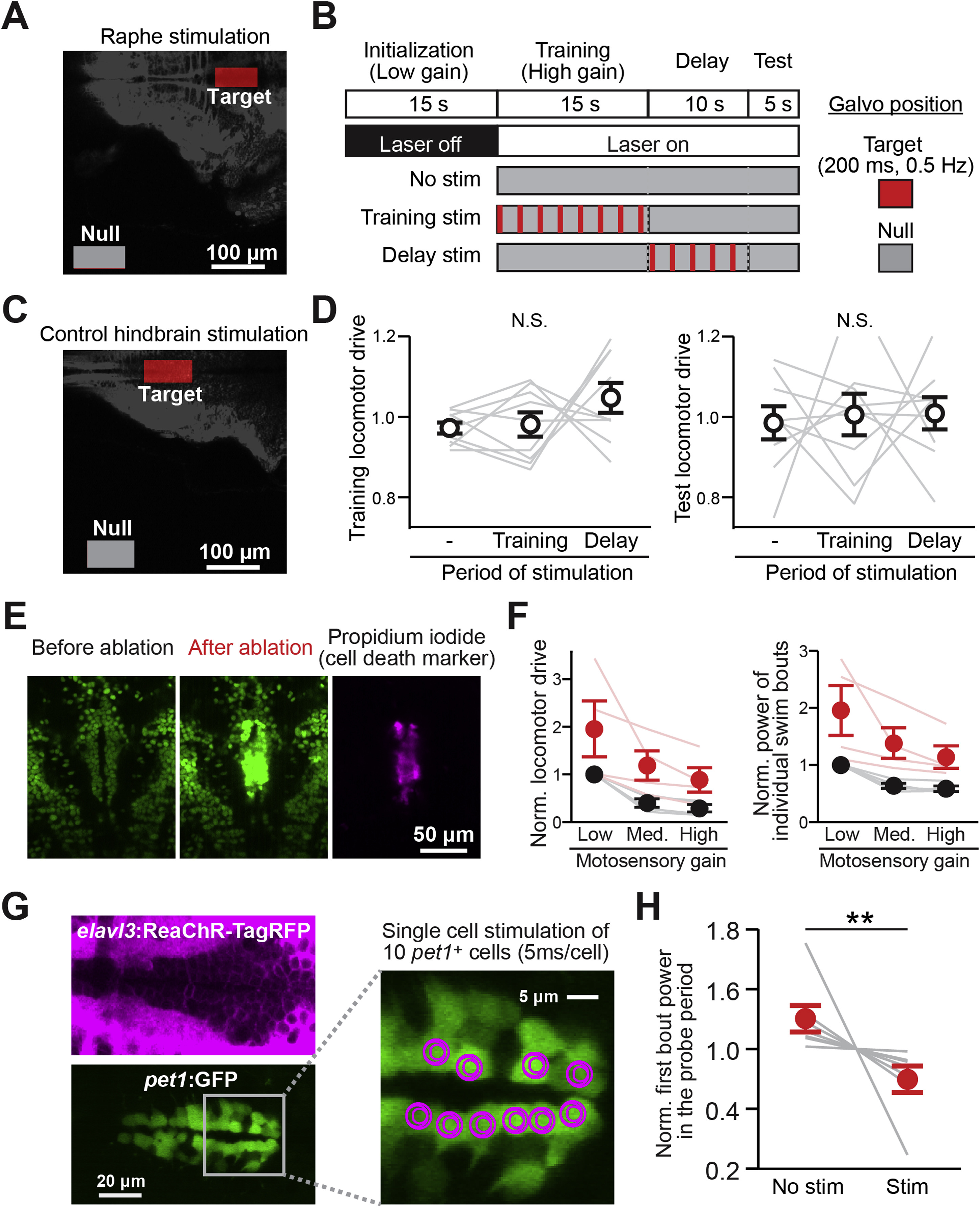
Optogenetic Activation and Laser Ablation of DRN Neurons, Related to Figure 6
(A) ROI settings for DRN stimulation. To stimulate DRN neurons, the target region is scanned, and to remove DRN stimulation, the null region is scanned (as an alternative to using a laser shutter, to avoid auditory stimulation by the rapid opening and closing of the shutter).
(B) Detailed stimulation protocol of the experiment presented in Figures 6E–6H. The default laser scan area was set to the ‘Null’ ROI outside the fish. During the period of stimulation, the laser scan area was moved to the ‘Target’ ROI for a short period of time (200 ms, performing four area scans of 50 ms each) once every 2 s.
(C) ROI settings for control hindbrain stimulation.
(D) Effect of control hindbrain stimulation on the locomotor drive in the training period (left) and the test period (right), showing no significant effects. N.S., p = 0.15 (left) and p = 0.92 (right) by one-way ANOVA across 9 fish. Error bars represent SEM across the same set of 9 fish as in Figures 6G and 6H. Gray lines represent data from individual fish.
(E) Two-photon plasma-mediated laser ablation of DRN neurons in Tg(elavl3:H2B-GCaMP6f)jf7 transgenic zebrafish. Dozens of cells in the DRN across multiple Z-planes were ablated; displayed here are images from a single plane. After ablation, the basal intensity of GCaMP fluorescence increased, indicating cell damage. Cell death and its local confinement were further verified post hoc by staining with propidium iodide, a membrane-impermeable dye-specific to dead cells.
(F) Locomotor drive (left) and power of individual swim bouts (right) under various levels of motosensory gain before (black) and after (red) DRN cell ablation, showing an increase in locomotor drive following ablation. The fish are still able to perform real-time motor adaptation, which we also observed in the chemical genetic ablation results in Figures 6 and S6. Values were normalized to the average locomotor drive under low motosensory gain before ablation in individual fish. Error bars represent SEM across 4 tested fish. Two-way ANOVA was performed on the effect of ablation and motosensory gain. (Left) p = 0.0045 and (Right) p = 0.0007 between before and after ablation. No interaction in the two-way ANOVA test was detected between the effects of ablation and motosensory gain. Thin red lines and gray lines represent data from individual fish before and after ablation, respectively.
(G) Cell-type specific two-photon optogenetic activation of individual serotonergic neurons in the DRN. Double transgenic zebrafish which express ReaChR-TagRFP under the elavl3 promoter (left top, magenta) and GFP in pet1+ serotonergic neurons (left bottom, green) [Tg(elavl3:ReaChR-TagRFP-T; pet1:GFP)] were used in this experiment. 10 pet1+ serotonergic neurons in the DRN were stimulated individually by local spiral scanning, with the scan pathway shown in magenta on the right. These neurons were stimulated 4 times in a 200 ms period (i.e., at 20 Hz within this window), with 5 ms spiral time per cell, timed to be 2 s before the end of the delay period (i.e., about 3 s before the first swim bout in the test period).
(H) Effect of stimulation during the delay period on the power of the first bout in the test period. The behavioral paradigm of this experiment is the same as the one in Figure 6F except that we only tested two conditions, control (No stim) and stimulation (Stim) as described in (G). ∗∗p = 0.0078 from one-tailed Wilcoxon signed-rank test across 7 fish for the effect of suppression after stimulation. Gray lines represent data from individual fish. Error bars: SEM across fish.
Reprinted from Cell, 167, Kawashima, T., Zwart, M.F., Yang, C.T., Mensh, B.D., Ahrens, M.B., The Serotonergic System Tracks the Outcomes of Actions to Mediate Short-Term Motor Learning, 933-946.e20, Copyright (2016) with permission from Elsevier. Full text @ Cell