
- Title
-
Inhibition of the TGFβ Pathway Enhances Retinal Regeneration in Adult Zebrafish
- Authors
- Tappeiner, C., Maurer, E., Sallin, P., Bise, T., Enzmann, V., Tschopp, M.
- Source
- Full text @ PLoS One

Immunohistological staining for P-Smad3 as an indicator of TGFβ pathway activation. The red channel with P-Smad3 staining is shown in the figures above, whereas overlay with the green (autofluorescence of photoreceptor outer segments) and blue channel (DAPI) is shown below. No relevant staining for P-Smad3 (red) was observed in the uninjured retina and one day after induction of retina degeneration with MNU. Starting at day 3 and until day 8, immunohistochemical staining for P-Smad3 revealed the activation of the TGFβ pathway (exemplarily, day 5 is shown). At day 15 and thereafter, no relevant activation was observed anymore (exemplarily, day 30 is shown). When the TGFβ pathway was inhibited (small molecule inhibitor SB431542), reduced staining for P-Smad3 was observed, when compared to the non-inhibited group in 0.1% dimethyl sulfoxide (DMSO). Lower magnification of retina 3 days after MNU treatment, including the peripheral retina is shown on the right. Cell nuclei are stained with DAPI (blue). The scale bar indicates 50 μm. GC: ganglion cells; INL: inner nuclear layer; ONL: outer nuclear layer. |
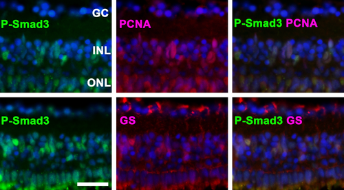
P-Smad3 is activated in proliferating cells. Top: The co-localization of P-Smad3 and proliferating cell nuclear antigen (PCNA) indicates that Smad3 is activated in proliferating cells. Bottom: P-Smad3-positive cells in the inner nuclear layer (INL) co-localized with glutamine synthetase (GS), suggesting that these cells are Müller glia. Representative immunohistochemical staining at day 3 is depicted. Cell nuclei are stained with DAPI (blue). The scale bar indicates 25 μm. GC: ganglion cells, ONL: outer nuclear layer. |
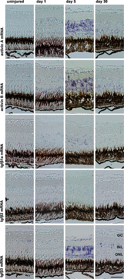
In situ hybridization with activin A and B as well as tgfβ1a, 2 and 3 antisense probes in zebrafish after the induction of retinal degeneration by MNU. Expression of these genes was detected beginning at day 1 and peaking at day 5. The highest staining intensity was observed for tgfβ3 and activins A and B, whereas only modest staining was observed for tgfβ1a and 2. These ligands were primarily detected in the inner nuclear layer (INL). The scale bar indicates 50 μm. GC: ganglion cells, ONL: outer nuclear layer. EXPRESSION / LABELING:
|

H&E staining of zebrafish retinas before (uninjured) and after induction of retina degeneration with MNU. In the non-inhibited (0.1% dimethyl sulfide, DMSO) and inhibited group (small molecule inhibitor SB431542), a reduction of rod cells was observed starting at day 3. In the non-inhibited group the reduction of rod photoreceptors persisted until day 8, whereas in the group with the inhibited TGFβ pathway (small molecule inhibitor SB431542) a rapid recovery was observed already at day 5. Scale bar indicates 50 μm. GC: ganglion cells, INL: inner nuclear layer, ONL: outer nuclear layer, SB: SB431542 PHENOTYPE:
|

Cell proliferation in the zebrafish retina exposed to 150 mg/l MNU. Proliferating cell nuclear antigen (PCNA) positive cells (red) indicate proliferation. Cell proliferation in the inner nuclear layer (INL) was highest at day 3 and 5, with no relevant difference between the non-inhibited (0.1% dimethyl sulfide, DMSO) and inhibited group (small molecule inhibitor SB431542). In contrast, proliferation in the outer nuclear layer (ONL) was higher in the inhibited group between 3 and 8. Cell nuclei are stained with DAPI (blue). Scale bar indicates 50 μm. GC: ganglion cells. PHENOTYPE:
|

TUNEL positive cells in the zebrafish retina after exposure to MNU. In uninjured zebrafish retina there are merely no TUNEL positive cells. Three days after exposure to 150 mg/l MNU, both the non-inhibited (0.1% dimethyl sulfide, DMSO) and the inhibited group (small molecule inhibitor SB431542) show a considerable amount of TUNEL positive cells (green) in the outer nuclear layer (ONL) and to a lesser degree in the inner nuclear layer (INL). Cell nuclei are stained with DAPI (blue). Scale bar indicates 50 μm. GC: ganglion cells. PHENOTYPE:
|