
Image
|
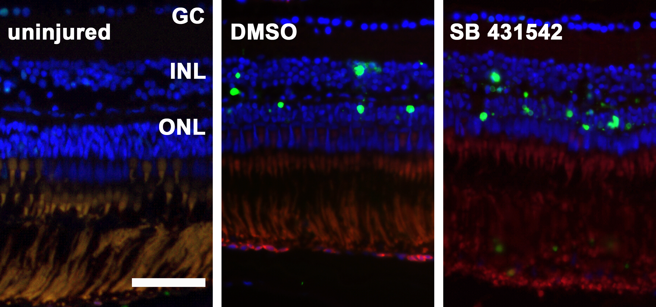
Figure Caption
Fig. 7
TUNEL positive cells in the zebrafish retina after exposure to MNU.
In uninjured zebrafish retina there are merely no TUNEL positive cells. Three days after exposure to 150 mg/l MNU, both the non-inhibited (0.1% dimethyl sulfide, DMSO) and the inhibited group (small molecule inhibitor SB431542) show a considerable amount of TUNEL positive cells (green) in the outer nuclear layer (ONL) and to a lesser degree in the inner nuclear layer (INL). Cell nuclei are stained with DAPI (blue). Scale bar indicates 50 μm. GC: ganglion cells.
Figure Data
Acknowledgments
This image is the copyrighted work of the attributed author or publisher, and
ZFIN has permission only to display this image to its users.
Additional permissions should be obtained from the applicable author or publisher of the image.
Full text @ PLoS One