
FIGURE
Fig. 2
- ID
- ZDB-FIG-161222-25
- Publication
- Tappeiner et al., 2016 - Inhibition of the TGFβ Pathway Enhances Retinal Regeneration in Adult Zebrafish
- Other Figures
- All Figure Page
- Back to All Figure Page
Fig. 2
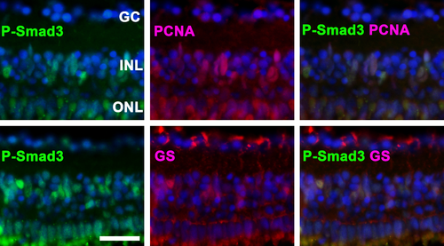
P-Smad3 is activated in proliferating cells. Top: The co-localization of P-Smad3 and proliferating cell nuclear antigen (PCNA) indicates that Smad3 is activated in proliferating cells. Bottom: P-Smad3-positive cells in the inner nuclear layer (INL) co-localized with glutamine synthetase (GS), suggesting that these cells are Müller glia. Representative immunohistochemical staining at day 3 is depicted. Cell nuclei are stained with DAPI (blue). The scale bar indicates 25 μm. GC: ganglion cells, ONL: outer nuclear layer. |
Expression Data
Expression Detail
Antibody Labeling
Phenotype Data
Phenotype Detail
Acknowledgments
This image is the copyrighted work of the attributed author or publisher, and
ZFIN has permission only to display this image to its users.
Additional permissions should be obtained from the applicable author or publisher of the image.
Full text @ PLoS One