- Title
-
Expression dynamics of NADPH oxidases during early zebrafish development
- Authors
- Weaver, C.J., Fai Leung, Y., Suter, D.M.
- Source
- Full text @ J. Comp. Neurol.

ZFIN is incorporating published figure images and captions as part of an ongoing project. Figures from some publications have not yet been curated, or are not available for display because of copyright restrictions. EXPRESSION / LABELING:
|
|
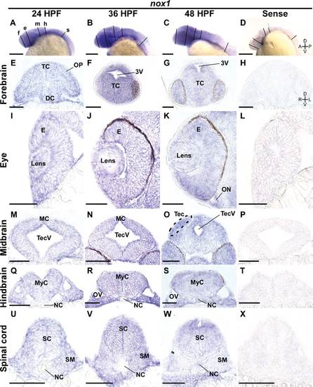
Broad nox1 expression through the first 2 days of development. A-D: Lateral views of whole-mount ISH embryos probed with antisense (A-C) and sense control (D) riboprobe against zebrafish nox1 mRNA. Lines represent the position of sections shown in E-X. E-H: 10-µm-thick transverse sections through the forebrain (line labeled “f” in A) of 24, 36, and 48 hpf embryos incubated with antisense probes (E-G, respectively) and 24 hpf embryo incubated with a sense control probe (H). I-L: Transverse sections through the eye (line labeled “e” in A). M-P: Corresponding midbrain sections (line labeled “m” in A). Q-T: Corresponding hindbrain sections (line labeled “h” in A). U-X: Corresponding spinal sections (line labeled “s” in A). Abbreviations: 3V, third ventricle; DC, diencephalon; E, eye; MC, mesencephalon; MyC, myelencephalon; NC, notochord; ON, optic nerve; OV, otic vesicle; SC, spinal cord; SM, somites; TC, telencephalon; Tec, tectum; TecV, tectal ventricle. Scale bar = 0.5 mm in A-D; 100 µm in E-X. |
|
Broad nox2/cybb expression through the first two days of development. A-D: Lateral views of whole-mount ISH embryos probed with antisense (A-C) and sense control (D) riboprobe against zebrafish nox2/cybb mRNA. Lines represent the position of sections shown in E-X. E-H: 10-µm-thick transverse sections through the forebrain (line labeled “f” in A) of 24, 36, and 48 hpf embryos incubated with antisense probes (E-G, respectively) and 24 hpf embryo probed with a sense control (H). I-L: Transverse sections through the eye (line labeled “e” in A). M-P: Corresponding midbrain sections (line labeled “m” in A). Q-T: Corresponding hindbrain sections (line labeled “h” in A). U-X: Corresponding spinal sections (line labeled “s” in A). Abbreviations: 3V, third ventricle; DC, diencephalon; E, eye; MC, mesencephalon; MyC, myelencephalon; NC, notochord; ON, optic nerve; OV, otic vesicle; SC, spinal cord; SM, somites; TC, telencephalon; Tec, tectum; TecV, tectal ventricle. Scale bar = 0.5 mm in A-D; 100 µm in E-X. |
|
Broad nox5 expression through the first 2 days of development. A-D: Lateral views of whole-mount ISH embryos probed with antisense (A-C) and sense control (D) riboprobe against zebrafish nox5 mRNA. Lines represent the position of sections shown in E-X. E-H: 10-µm-thick transverse sections through the forebrain (line labeled “f” in A) of 24, 36, and 48 hpf embryos incubated with antisense probes (E-G, respectively) and 24 hpf embryo probed with a sense control (H). I-L: Transverse sections through the eye (line labeled “e” in A). M-P: Corresponding midbrain sections (line labeled “m” in A). Q-T: Corresponding hindbrain sections (line labeled “h” in A). U-X:. Corresponding spinal sections (line labeled “s” in A). Abbreviations: 3V, third ventricle; DC, diencephalon; E, eye; MC, mesencephalon; MyC, myelencephalon; NC, notochord; ON, optic nerve; OV, otic vesicle; SC, spinal cord; SM, somites; TC, telencephalon; Tec, tectum; TecV, tectal ventricle. Scale bar = 0.5 mm in A-D; 100 µm in E-X. |
|
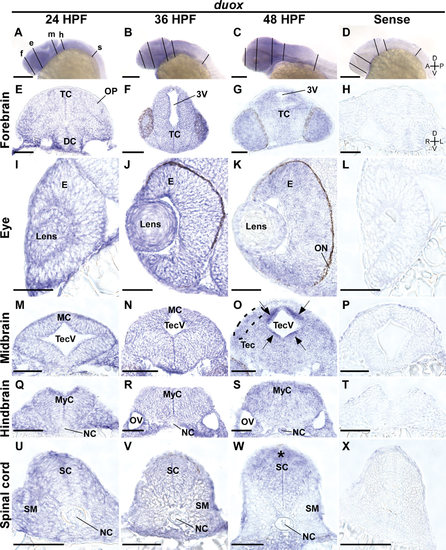
duox is highly expressed around tectal ventricle at 48 hpf . A-D: Lateral views of whole-mount ISH embryos probed with antisense (A-C) and sense control (D) riboprobe against zebrafish duox mRNA. Lines represent the position of sections shown in E-X. E-H: 10-µm-thick transverse sections through the forebrain (line labeled “f” in A) of 24, 36, and 48 hpf embryos incubated with antisense probes (E-G, respectively) and 24 hpf embryo probed with a sense control (H). I-L: Transverse sections through the eye (line labeled “e” in A). M-P: Corresponding midbrain sections (line labeled “m” in A). High duox expression was detected around the tectal ventricle (arrows in O). Q-T: Corresponding hindbrain sections (line labeled “h” in A). U-X: Corresponding spinal sections (line labeled “s” in A). Asterisk and arrows represent regions and small areas of increased duox expression in the spinal cord and midbrain, respectively. Abbreviations: 3V, third ventricle; DC, diencephalon; E, eye; MC, mesencephalon; MyC, myelencephalon; NC, notochord; ON, optic nerve; OV, otic vesicle; SC, spinal cord; SM, somites; TC, telencephalon; Tec, tectum; TecV, tectal ventricle. Scale bar = 0.5 mm in A-D; 100 µm in E-X. |


