- Title
-
Sfrp5 Modulates Both Wnt and BMP Signaling and Regulates Gastrointestinal Organogensis in the Zebrafish, Danio rerio
- Authors
- Stuckenholz, C., Lu, L., Thakur, P.C., Choi, T.Y., Shin, D., and Bahary, N.
- Source
- Full text @ PLoS One
|
Expression profile of sfrp5. Expression level of sfrp5 as measured by probeset Dr.21012.1.S1 in GI tissue (dark green squares) and non-GI tissue (light green triangles) from 2 through 6 dpf (for details, see [39]). B) Expression of sfrp5 and β-actin by RT-PCR of total RNA isolated at indicated time points. C–O) Whole-mount in situ hybridization showing sfrp5 expression in zebrafish embryos at 3 hpf (C), shield stage (D), 8 hpf (E), bud stage (F), early (G), mid (H), and late somitogenesis (I), 24 hpf (J), 32 hpf (K), 2 dpf (L), 3 dpf (M), 4 dpf (N), and 6 dpf (O). Lateral views with animal pole to the top (C–F) or with anterior to the left (G–I, M–O). Dorsal view with anterior to the left (J–L). |
|
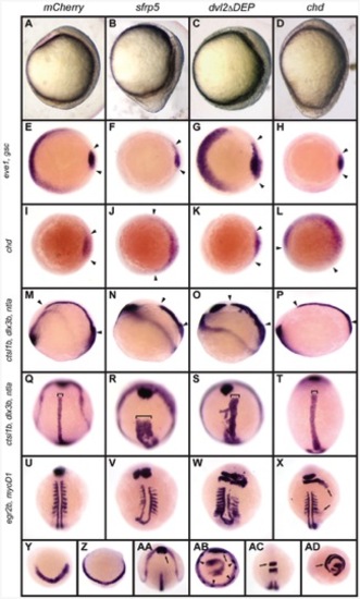
Overexpression of sfrp5 disrupts gastrulation. Embryos were injected with 100 pg mCherry as control (A, E, I, M, Q, U, Y, AA, AC), 140 pg sfrp5 (B, F, J, N, R, V, Z, AB, AD), 150 pg dvl2ΔDEP (C, G, K, O, S, W), or 50 pg chd (D, H, L, P, T, X). A–D) Morphology of injected embryos injected at early somitogenesis, lateral view with dorsal side to right. E–AD) Whole-mount in situ hybridization of injected embryos. E–H) Animal pole view with dorsal side to the right of embryos stained with eve1 and gsc probes at shield stage. Arrowheads demarcate gsc staining. I–L) Animal pole view with dorsal side to the right of embryos stained with chd, demarcated by arrowheads, at shield stage. M–T) Early somitogenesis embryos stained with probes against ctsl1b, dlx3b, and ntla. M–P: Lateral view with dorsal to top. Arrowheads mark length of notochord. Q–T: dorsal view with anterior to top. Brackets show notochord width. U–X) Mid-somitogenesis embryos stained with egr2b and myoD1. Dorsal view with anterior to top. In X, arrows mark radialization of egfr2b and myoD1 staining. Y, Z) Bud-stage embryos hybridized with probe against her5; anterior view, dorsal to bottom. AA–AB) Early somitogenesis embryos hybridized with probes against ctsl1b, dlx3b, and ntla. Arrow: normal ctsl1b staining. Arrowhead: ectopic ctsl1b staining. AA: Dorsal view, anterior to top. AB: Ventral view, dorsal to top. AC–AD) Mid-somitogenesis embryos hybridized with probes against egr2b and myoD1. AC: Dorsal view, anterior to top. AD: ventral view, dorsal to top. Arrows point to rhombomere 3. |
|
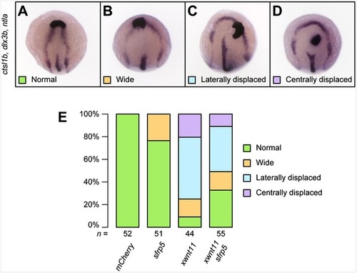
sfrp5 overexpression inhibits non-canonical Wnt signaling. Embryos injected with 200 pg mCherry, 140 pg sfrp5, and 100 pg wnt11b from Xenopus laevis alone and in combination as indicated. A–D) 4-somite embryos were processed by in situ hybridization with probes against ctsl1b, dlx3b, and ntla. Dorsal view with anterior towards the top. The neural plate was scored as normal (A), widened (B), or widened with a laterally (C) or centrally (D) displaced prechordal plate. E) Bar chart showing percentage of different phenotypic classes for each treatment. The total number of embryos analyzed per treatment is shown under each column. |
|
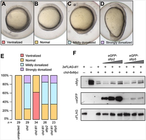
Sfrp5 inhibits the Tll1 protease. Embryos were injected with combinations of 50 pg chd, 200 pg tll1, 150 pg sfrp2, and 125 pg sfrp5. At the 4 somite stage, embryos were classified as ventralized (A), normal (B), mildly dorsalized (corresponding to C1– C3; C) or strongly dorsalized (C4– C5; D) [59] and the results plotted (E). The numbers under each bar represent the number of embryos analyzed per treatment. F) Conditioned media from singly transfected 293T cells were combined as indicated, incubated, and analyzed by Western blotting. Volume of conditioned media from tll1 and chd transfected cells was kept constant when used, but volume of conditioned media from sfrp2 or sfrp5 transfected cells was doubled or tripled as indicated. |
|
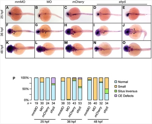
Sfrp5 regulates hepatoblast formation in zebrafish. Embryos were injected with 0.5 pmol morpholino against sfrp5 (MO; B, G, L, Q), 0.5 pmol of the control morpholino (mmMO; A, F, K, P), 100 pg mCherry mRNA (C, H, M, R), or 100 pg sfrp5 mRNA (D, E, I, J, N, O, S, T). A–O) Whole-mount in situ hybridization with a probe against hhex staining embryos at 25 hpf (A–E), 36 hpf (F–J), and 48 hpf (K–O). Dorsal view, anterior to the left. Arrowheads point to the hepatoblast. P–T) Confocal microscopy of injected gutGFP embryos. Ventral view with anterior to top. Liver is outlined in white, pancreas in yellow. The scale bar is equal to 25 μm. U) RT-PCR of morpholino and control injected embryos with primer pairs detecting sfrp5 or β-actin. V) Bar chart representing distribution of normal and abnormal embryos processed by in situ hybridization, with representative samples shown in A–O. W) Boxplot showing liver size distribution in injected embryos. **: p<0.001. X) Boxplot showing distribution of the number of GFP+ liver cells. *: p<0.05, **: p<0.01. Y) Boxplot showing liver cell size distribution. Numbers below each column or boxplot show how many embryos were analyzed. |
|
Endodermal defects in embryos with altered levels of Sfrp5. Embryos were injected with 0.5 pmol morpholino against sfrp5 (MO; panels B, G, L), 0.5 pmol of the control morpholino (mmMO; panels A, F, K), 100 pg mCherry mRNA (C, H, M), or 100 pg sfrp5 mRNA (D, E, I, J, N, O). A–O) Whole-mount in situ hybridization with a probe against foxa1 staining embryos at 25 hpf (A–E), 36 hpf (F–J), and 48 hpf (K–O). Dorsal view, anterior to the left. Arrowheads point to the hepatoblast (F–J). P) Bar chart representing distribution of normal and abnormal embryos processed by in situ hybridization, with representative samples shown in A–O. Below each column, numbers of embryos analyzed. |
|
Modulation of sfrp5 expression causes defects in gastrointestinal development. A–P) Dorsal views of 3 dpf old embryos stained with probes against fabp10a (A–D), anxa2b (E–H), try (I–L), and ins (M–O). Embryos were injected with 0.5 pmol mismatch morpholino (A, E, I, M), 0.5 pmol morpholino against sfrp5 (B, F, J, N), 100 pg eGFP mRNA (C, G, K, O) or 50 pg sfrp5 mRNA (D, H, L, P). Q) Chart summarizing in situ results (A–L), showing percentages of normal or small GI organs. R) Chart summarizing in situ results (M–P), showing the percentage of larvae in which ins+ cells failed to coalesce. The total number of analyzed embryos per treatment is shown below each column. EXPRESSION / LABELING:
PHENOTYPE:
|
|
Overexpression of sfrp5 inhibits canonical Wnt signaling. Clutches of 1- to 2-cell stage embryos obtained from an outcross of Tg(hs:mCherry,wnt2bb) heterozygotes with wild type fish were injected with either 100 pg eGFP or 100 pg sfrp5 and sorted based on expression of mCherry after heat shock. A-D) 48 hpf embryos were analyzed for liver formation by in situ hybridization with hhex and categorized as having an enlarged (A), normal (B), or small liver (C). We also observed some embryos without apparent hepatoblast (D) or in which the hepatoblast failed to coalesce into a single field (E). Square brackets indicate the size of the hepatoblast, the asterisk in (D) mislocalized hhex positive cells. F) Bar chart showing the percentages of each category per treatment. The total number of analyzed embryos per treatment is shown below each column. EXPRESSION / LABELING:
|
|
The notochord undulates and is kinked in sfrp5 and dvl2ΔDEP injected embryos. All embryos were processed by in situ hybridization using a cocktail of probes against ctsl1b, dlx3b, and ntla and are shown in dorsal view, anterior to top. A) Embryo injected with 200 pg of mCherry mRNA. B) Embryo injected with 50 pg sfrp5 mRNA. C) Embryo injected with 140 pg sfrp5 mRNA. D) Embryo injected with 150 pg dvl2ΔDEP mRNA. Arrows point to the notochord. |


