
- Title
-
Functional modeling in zebrafish demonstrates that the atrial-fibrillation-associated gene GREM2 regulates cardiac laterality, cardiomyocyte differentiation and atrial rhythm
- Authors
- Müller, I.I., Melville, D.B., Tanwar, V., Rybski, W.M., Mukherjee, A., Shoemaker, B.M., Wang, W.D., Schoenhard, J.A., Roden, D.M., Darbar, D., Knapik, E.W., and Hatzopoulos, A.K.
- Source
- Full text @ Dis. Model. Mech.
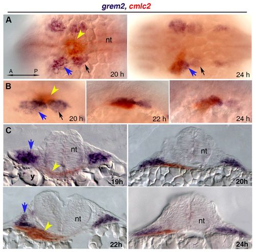
Expression ofgrem2 in pharyngeal arch primordia during cardiac tube migration. (A,B) Whole mount images of grem2 (purple)- and cmlc2 (red)-stained embryos in dorsal (A) and lateral (B) views, anterior to the left. grem2 is expressed in the first two pharyngeal arches (blue arrows point to the left first arch, black arrows to the left second arch) as the cardiac progenitors begin to assemble in the midline. During cardiac jogging to the left, the heart tube (yellow arrowhead) is positioned next to grem2-expressing pharyngeal mesoderm and subsequently passes ventrally to the grem2-expression domain in the left first pharyngeal arch. (C) Cross-sections of zebrafish embryos double-labeled for grem2 and cmlc2. h, hours post fertilization; A, anterior; P, posterior; nt, neural tube; y, yolk. EXPRESSION / LABELING:
|
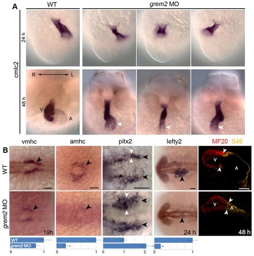
Loss of Grem2 leads to cardiac jogging and looping defects, aberrant expression ofpitx2and abnormal development of cardiac chambers. (A) In situ hybridization analysis using cmlc2 riboprobe shows that in wild-type (WT) embryos the heart jogs leftward, whereas cardiac morphogenesis is randomized in grem2 morphants (grem2 MO). The cardiac tube, consisting of a single atrium (A, white arrowheads) and a single ventricle (V), is thinner, shorter and fails to loop in grem2 morphants. Left/Right (L/R) axis orientation is indicated. (B) In situ hybridization analysis using vmhc and amhc riboprobes shows that loss of Grem2 leads to lower expression levels of both vmhc and amhc, with amhc being essentially absent at 19 hpf (arrowheads). Pitx2 expression (black arrowheads) at 19 hpf is enhanced (white arrowheads), whereas lefty2 expression at 24 hpf is abolished (black arrowheads). Quantification of expression levels in wild-type and morphant embryos is shown below the corresponding images (expression is relative to wild type, which was set as arbitrary value 1). Immunofluorescence analysis using MF20 (red, labels both ventricular and atrial cardiomyocytes) and S46 (green, labels only atrial cardiomyocytes, which appear yellow) antibodies shows abnormal development of both chambers, with the atrium being more deformed and reduced in size than the ventricle. Arrowheads mark the position of the atrio-ventricular boundary. Scale bars: 50 μm. *P<0.05. EXPRESSION / LABELING:
|
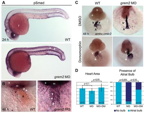
Loss of Grem2 leads to increased BMP signaling and cardiac defects that can be reversed by the BMP inhibitor dorsomorphin. (A) Whole mount immunohistochemistry using antibodies recognizing the phosphorylated forms of Smads1/5/8 shows stronger pSmad protein staining in grem2 morphants than in controls. Arrowhead marks somite boundaries. (B) Frontal close up views of the heart after pSmad1/5/8 antibody staining at 48 hpf shows sharply increased nuclear staining in cardiomyocytes of Grem2-depleted embryos. The heart is outlined by dotted lines; arrowheads mark pSmad-positive nuclei. (C) Wild-type (WT) embryos and grem2 morphants were incubated with dorsomorphin or its vehicle DMSO between 16 and 48 hpf and stained at 48 hpf with cmlc2 and amhc probes to visualize the ventricle (V) and atrium (A). Dorsomorphin treatment restored atrial patterning and differentiation (arrowheads point to the atrioventricular boundary in wild-type hearts; dotted lines demarcate ventricle-atrium boundary in grem2 morphants). (D) Quantification of heart size (Heart Area) and atrial bulb restoration (Presence of Atrial Bulb) in DMSO-treated controls (WT), DMSO-treated grem2 morphants (MO) and grem2 morphants treated with dorsomorphin (MO+DM). Dorsomorphin treatment restores atrial development. Error bars represent s.d. P-values and number of embryos analyzed (n) are indicated. b, brain; e, eye; m, mouth; y, yolk. |
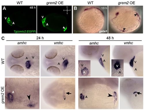
Overexpression of Grem2 induces ectopic atrial myocardium. (A) Lateral view of live wild-type (WT) and grem2 mRNA-injected (15 pg) cmlc2-egfp transgenic embryos (grem2 OE) at 48 hpf shows ectopic cardiac tissue (arrowhead). Anterior-posterior (A/P) and dorsal-ventral (D/V) axes are indicated. (B) In situ hybridization shows ectopic sites expressing gata-5 along the paraxial mesoderm (arrowheads). (C) WT zebrafish embryos were injected with grem2 mRNA at the 1-4 cell stage; non-injected embryos served as controls. At 24 hpf, Grem2 overexpression (OE) led to enlarged or ectopic (arrowhead) amhc expression in 100% of injected embryos, whereas vmhc expression was reduced (arrow) or absent in 54.4%. These results remained consistent at 48 hpf, with 96% of embryos displaying ectopic amhc expression (arrowhead), and 52.2% displaying reduced vmhc expression (arrow). Insets display enlarged frontal images. Normal atrium (A) expressing amhc and ventricle (V) expressing vmhc are marked; dorsal (24 hpf) and lateral (48 hpf) views; anterior is to the left in all panels. |
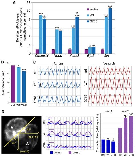
Overexpression of Grem2 induces ectopic atrial myocardium. (A) Lateral view of live wild-type (WT) and grem2 mRNA-injected (15 pg) cmlc2-egfp transgenic embryos (grem2 OE) at 48 hpf shows ectopic cardiac tissue (arrowhead). Anterior-posterior (A/P) and dorsal-ventral (D/V) axes are indicated. (B) In situ hybridization shows ectopic sites expressing gata-5 along the paraxial mesoderm (arrowheads). (C) WT zebrafish embryos were injected with grem2 mRNA at the 1-4 cell stage; non-injected embryos served as controls. At 24 hpf, Grem2 overexpression (OE) led to enlarged or ectopic (arrowhead) amhc expression in 100% of injected embryos, whereas vmhc expression was reduced (arrow) or absent in 54.4%. These results remained consistent at 48 hpf, with 96% of embryos displaying ectopic amhc expression (arrowhead), and 52.2% displaying reduced vmhc expression (arrow). Insets display enlarged frontal images. Normal atrium (A) expressing amhc and ventricle (V) expressing vmhc are marked; dorsal (24 hpf) and lateral (48 hpf) views; anterior is to the left in all panels. |
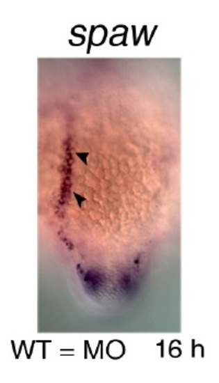
Loss of Grem2 does not affect early asymmetric gene expression of the nodal gene spaw in posterior mesoderm. Spaw expression at 16 hours post-fertilization (h) in grem2 morphants is indistinguishable from wild-types. |