- Title
-
Zebrafish foxP2 Zinc Finger Nuclease Mutant Has Normal Axon Pathfinding
- Authors
- Xing, L., Hoshijima, K., Grunwald, D.J., Fujimoto, E., Quist, T.S., Sneddon, J., Chien, C.B., Stevenson, T.J., and Bonkowsky, J.L.
- Source
- Full text @ PLoS One
|
Flow diagram for generation of ZFN mutants, and HRMA analysis. (A) Flow diagram illustrating steps in generation and identification of fish carrying ZFN-induced mutation. (B) Illustrative melt-curve (“HRMA” high-resolution melt analysis) of F1 generation foxP2 ZFN fish. Y-axis corresponds to the differential of the change (decrease) in fluorescence; X-axis is the temperature (°C). Each colored line is a different sample, arrows point to samples with mutations in the foxP2 amplicon; asterisk is the wild-type product. There is some variability in the melt temperature of the wild-type amplicon because of minor variations in starting template amount and salt concentrations. (C-C2) Melt-curves of dilutions of mutant foxP2 DNA. Abnormal melt curves (arrowheads) are detected at dilutions of 1:20 mutant:wild-type DNA, but not (or very minimally) at a dilution of 1:50. (D) Schematic diagram of foxP2 protein domains; asterisk shows the targeted region for ZFN mutagenesis; sequence above the picture is the nucleotide sequence targeted. Below are shown the three alleles, with the sequence of the mutation, and a picture of the predicted protein. (E) RT-PCR from wt or foxP2 homozygous mutant embryos at 72hpf, with primers for the full-length transcript, or encompassing exons 1–2. No alternative splice variants were noted, and sequencing of the foxP2 mutant PCR product showed that the mutations led to the predicted out-of-frame sequence. EXPRESSION / LABELING:
|
|
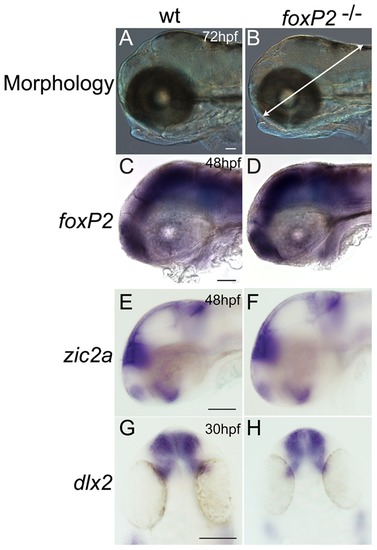
foxP2 mutant has normal morphology and CNS patterning. Whole-mount embryos, brightfield images, scale bar = 50 μm. Lateral views, rostral to the left (A-F); dorsal views, rostral to the top (G, H). (A, B) Gross head morphology is unchanged in mutants at 72hpf. Arrow in (B) shows line used to measure brain size, from the midbrain-hindbrain boundary to the edge of the head by dissecting the midpoint of the lens. (C-D) mRNA expression of foxP2 transcript is unchanged in intensity and pattern in foxP2 mutants. (E-H) in situ expression patterns and intensity of zic2a and dlx2 is unchanged in mutants compared to wild-type embryos. PHENOTYPE:
|
|
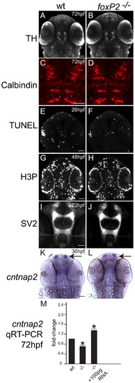
foxP2 does not affect neuron specification, apoptosis, or proliferation, but does affect cntnap2 expression levels. Confocal z-stack images, rostral to the top, scale bars 50 μm. (A, B, E-H, K-L) ventral views. (C, D) dorsal views. (A, B) TH immunohistochemistry at 72hpf shows no difference in WT and mutants in diencephalic dopaminergic neuron pattern or number. (C, D) Calbindin immunohistochemistry in the dorsal hindbrain at 72hpf shows similar patterns and numbers of Purkinje neurons in WT and mutants. (E, F) TUNEL staining for apoptotic cells at 26hpf in the brain shows no difference in pattern or number of cells between WT and mutants. (G, H) Detection of proliferation by H3P staining is similar in WT and mutants at 48hpf. (I, J) Neuropil distribution and intensity visualized with anti-SV2 synaptic vesicle protein antibody at 72hpf is similar in WT and mutants. (K, L) cntnap2 in situ expression pattern at 36hpf shows less expression in mutant embryos, more noticeably in the telencephalon (arrows). (M) quantitative RT-PCR at 72hpf confirms decreased expression of cntnap2 in mutants, which is rescued by injection with full-length foxP2 mRNA (* p<0.05). Y-axis indicates fold-change. EXPRESSION / LABELING:
PHENOTYPE:
|
|
foxP2 does not affect axon pathfinding. Confocal z-stack images of whole-mount embryos, scale bars 50 μm, show no difference between wild-type and foxP2 mutant (/) embryos for axon pathfinding using a variety of axonal labels. (A–D) anti-SV2 immunohistochemistry at 24hpf, lateral views of the brain, rostral to the left (A, B) and 72hpf, dorsal views of the spinal cord, rostral to the top (C, D). (E, F) anti-acetylated tubulin immunohistochemistry at 72hpf, ventral views of the optic chiasm. (G-L) GFP immunohistochemistry at 72hpf in Tg(foxP2-enhancerA.2:egfp-caax) embryos that labels foxP2 neurons show no pathfinding errors in anterior commissure (ac), longitudinal commissures (lc), tract of the commissure of the posterior tuberculum (TCPTc), or reticulospinal axons (rs). (M-R) GFP immunohistochemistry at 72hpf in Tg(otpb.A:egfp-caax) embryos for visualization of dopaminergic and neuroendocrine projections (M, N, with insets shown in O, P) in the brain, and dopaminergic axon tracts in spinal cord (Q, R). EXPRESSION / LABELING:
|


