- Title
-
Diverse mechanisms for assembly of branchiomeric nerves
- Authors
- Cox, J.A., Lamora, A., Johnson, S.L., and Voigt, M.M.
- Source
- Full text @ Dev. Biol.
|
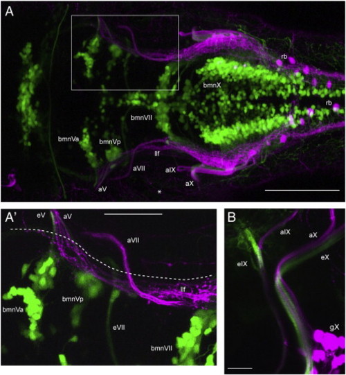
Cranial nerves V, IX and X contain both motor and sensory axons whereas the proximal VIIth is separated into motor or sensory alone. Images shown are confocal z-stacks obtained from 4 dpf larvae in which bmn neurons/axons are green and peripheral sensory axons are magenta. A shows a dorsal view (anterior to left) of a typical 4 dpf isl:gfp;3.2:cherry larva (n = 25). A′ is a higher power image from the same larva (boxed region in A) showing a dorsal view of the motor portion of the VIIth nerve as it exits the hindbrain below the descending trigeminal projections (llf), and of the sensory portion of the VIIth as it enters the hindbrain above the llf. The dashed line marks the approximate position of the hindbrain boundary. B is a lateral view (anterior to left, dorsal at top) caudal to the ear of a typical 4 dpf nkx:mgfp;3.2:cherry larva (nkx:mgfp was used as it contains labeling of bmnIX neurons, unlike the isl:gfp line; n = 7). It shows that the IXth and Xth cranial nerves contain both sensory and motor components at the hindbrain nerve root. This image also shows that the sensory and motor axons are present as separate fascicles within the nerves. bmnVa, anterior motor nucleus of V; bmnVp, posterior motor nucleus of V; bmnVII, motor nucleus of VII; eV, efferent nerve of bmnV; eVII, efferent nerve of bmnVII; eIX, efferent of bmnIX; eX, efferent nerve of bmnX; aV, afferent nerve of gV; aVII, afferent nerve of gVII; aIX, afferent nerve of gIX; aX, afferent nerve of gX; llf, lateral longitudinal fascicle; rb, Rohon–Beard neuron (in magenta); asterisk, peripheral processes of gV and RB neurons. Scale bars; A: 100 μm, A′: 50 μm, B: 20 μm. EXPRESSION / LABELING:
|
|
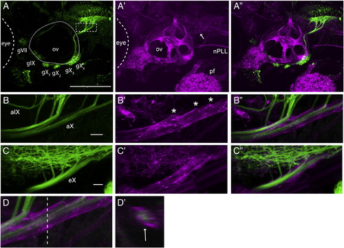
Cranial nerves containing sensory and motor axons are ensheathed by glia by 4 dpf. A shows a representative lateral view of GFP-expressing afferent axons in a 3.2:gfp;sox:rfp larva (n = 17), A′ shows RFP-expressing cells in the same larva (the arrow indicates the position of the vagal nerve sheath), and A" is a merge of A and A′. B is a higher power of the area indicted by the dashed rectangle in A showing GFP+ vagal afferents entering the hindbrain in a 3.2:gfp;sox:rfp larva; B′ shows the RFP+ glial sheath; B" is a merge of B and B′. Asterisks mark openings in the glial sheath where the vagal afferents leave the sheath to enter the hindbrain proper. C shows a lateral view of the bmnX efferents exiting the hindbrain in a 4 dpf nkx:mgfp;sox:rfp larva (n = 5) (also seen in green are oligodendrocyte processes in the hindbrain); C′ shows the glial sheath and C" is a merge of C and C′. D is a lateral view of aX in a 3.2:gfp;sox:rfp larva, D′ is an orthogonal projection (cross-section), taken at the level of the dotted line in D, showing the ensheathment of the peripheral portion of aX (indicated by arrow). gVII–gX, sensory ganglia; ov, otic vesicle; nPLL posterior lateral line nerve; aIX and aX- afferent nerves of gIX and gX; eX- efferent nerve of bmnX, pf, pectoral fin. All images are lateral, with anterior to left, dorsal at top. Scale bars: A: 100 μm, B: 20 μm, C: 20 μm. EXPRESSION / LABELING:
|
|
Sox:rfp+ peripheral glia contribute to the hindbrain transition zone of the Xth nerve. Images shown are from a 4 dpf olig2:gfp;sox:rfp larva (n = 3). Panel A is a dorsal view (anterior at left) of one side of the hindbrain, caudal to the otocyst, showing GFP+ oligodendrocytes and their processes. Arrows point to oligodendrocyte cell bodies and an arrowhead indicates an olig2-expressing cell present outside of the hindbrain. The dashed line marks the approximate position of the hindbrain boundary. A′ shows membrane-targeted RFP in the peripheral glia composing the vagal sheath, as well as in some CNS oligodendrocytes and their processes. Note the RFP+ processes (marked by asterisks) that appear to link the glial sheath to the hindbrain at several points. A" is a merge of A and A′. Notice that the GFP+ cell seen in panel A is present in the glial sheath. sm, skeletal muscle; gsX, glial sheath of the Xth nerve. Scale bars are 20 μm. EXPRESSION / LABELING:
|
|
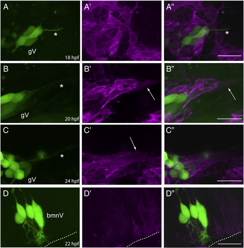
Sensory axons, followed by peripheral glia, are added to the Vth nerve prior to the addition of bmnV efferents. Row A: lateral view of a 3.2:gfp;sox:rfp embryo at 18 hpf (n = 4). The GFP channel (A) shows that the central afferents (marked by the asterisk) from trigeminal ganglion (gV) sensory neurons are growing toward their hindbrain targets, while the RFP (A′) and merged (A") channels show that there are no peripheral glia ensheathing the axon fascicle at this time point. Row B: lateral view of a 3.2:gfp;sox:rfp embryo at 20 hpf (n = 3). The GFP channel (B) shows the central afferents (marked by the asterisk) of the gV sensory neurons as they have extended further toward their hindbrain targets, while the RFP (B′) and merged (B") channels show that the peripheral glia have begun to ensheath the axon fascicle. The arrow in B′ and B" points to the glial sheath that is beginning to form. Row C: lateral view of a 3.2:gfp;sox:rfp embryo at 24 hpf (n = 11). The GFP channel (C) again shows the central afferents (marked by the asterisk) of the gV sensory neurons; the RFP (C′) and merged (C") channels show a well-developed glial sheath surrounding the sensory afferents. The arrow in C′ points to the glial sheath of the Vth nerve. Row D: lateral view of a isl:gfp;sox:rfp embryo at 22 hpf (n = 4). The GFP channel (D) shows that neurons within the Vth motor nucleus are just beginning to extend their efferents; the RFP (D′) and merged (D") channels show that these efferents have not yet extended from the hindbrain into the periphery. The dotted line indicates the margin of the hindbrain. In all images, anterior is to the left and dorsal at the top. Scale bars are 25 μm. EXPRESSION / LABELING:
|
|
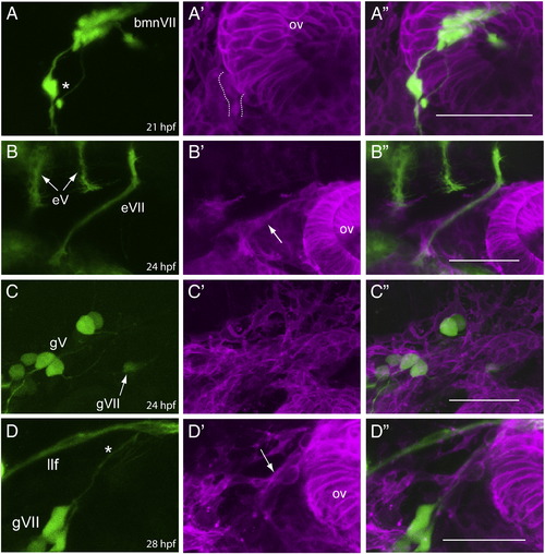
bmnVII axons enter into the periphery and become ensheathed by glia prior to the time when gVII afferents are formed and ensheathed. Row A: lateral view of an isl:gfp;sox:rfp embryo at 21 hpf (n = 6). The GFP channel (A) shows that the VII motor neurons (bmnVII) efferents (marked by the asterisk) are growing toward their peripheral targets, while the RFP (A′) and merged (A") channels show that RFP+ cells are associating with these axons at this time point (dotted lines indicate nerve-associated RFP+ cells). Row B: lateral view of an nkx:mgfp;sox:rfp embryo at 24 hpf (n = 3). The GFP channel (B) shows that the bmnVII axons have grown further in to the periphery, while the RFP (B′) and merged (B") channels show a definitive glial sheath (arrow in B′) has formed around the axon fascicle by this time. Also shown are the processes (marked eV) from the two bmnV nuclei at this stage which are still within the hindbrain, and have not yet joined to form the one efferent bundle that exits the hindbrain at r2. Row C: lateral view of a 3.2:gfp;sox:rfp embryo at 24 hpf (n = 8). The GFP channel (C) shows the gVII sensory neurons (marked by the arrow) are just beginning to form; the RFP (C′) and merged (C") channels show no discernable RFP+ cells are associated with these nascent neurons. Row D: lateral view of a 3.2:gfp;sox:rfp embryo at 28 hpf (n = 5). The GFP channel (D) shows that the afferent VII fibers (marked by the asterisk) have reached the region of the hindbrain margin; the RFP (D′) and merged (D") channels show that these afferents are now ensheathed by peripheral glia (marked by the arrow in D′). gV, trigeminal sensory ganglion neurons; llf, lateral longitudinal fascicle; ov, otic vesicle. In all images, anterior is to the left and dorsal at the top. Scale bars are 50 μm. EXPRESSION / LABELING:
|
|
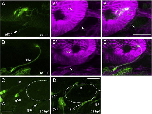
Formation of the IXth nerve is initiated by bmnIX efferents, which are followed by glia and then the gIX afferents. Row A: lateral view of a nkx:mgfp;sox:rfp embryo at 25 hpf (n = 4). The GFP channel (A) shows that the motor efferents from the bmnIX have just entered the periphery at this time, and the arrows in the RFP (A′) and merged (A") channels point to RFP+ peripheral glia that are associating with these axons. Row B: lateral view of a nkx:mgfp;sox:rfp embryo at 30 hpf (n = 5). The GFP channel (B) shows a well-defined efferent fascicle that has just reached the branchial arches, while the RFP (B′) and merged (B") channels show that a definitive sheath (marked by the arrow in B) has encompassed this fascicle. Panel C: lateral view of a 3.2:gfp embryo at 32 hpf shows that the first neurons forming the gIX are just appearing at this time (n = 12). Panel D: lateral view of a 3.2:gfp embryo at 38 hpf shows that the gIX afferents (marked by the asterisk) are just reaching the margin of the hindbrain (n = 11). The dotted line indicates the position of the otic vesicle. gV, trigeminal ganglion sensory neurons; gVII, facial ganglion sensory neurons; gX, vagal ganglion sensory neurons. In all images, anterior is to the left and dorsal at the top. Scale bars are 25 μm. |
|
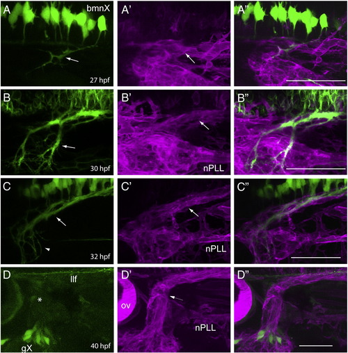
Formation of the Xth nerve is initiated by bmnX efferents, which are followed by glia and then the gX afferents. Row A: lateral view of a isl:gfp;sox:rfp embryo at 27 hpf (n = 5). The GFP channel (A) shows that the vagal motor (bmnX) efferents (marked by the arrow) have entered the periphery, while the RFP (A′) and merged (A") channels show that peripheral glia are associating with the axon fascicles at this time point. Row B: lateral view of a isl:gfp;sox:rfp embryo at 30 hpf (n = 3). The GFP channel (B) shows the bmnX efferents (marked by the arrow) are present as several fascicles, and the RFP (B′) and merged (B") channels show that each set of axons is now enclosed by a defined glial sheath (arrow in B′). Row C: lateral view of a isl:gfp;sox:rfp embryo at 32 hpf (n = 3). The GFP channel (C) shows that the more proximal vagal efferent axons are coalescing to form a distinct fascicle (arrow) whereas the growth cone field of these axons remains diffuse (arrowhead); the RFP (C′) and merged (C") channels show a well-developed glial sheath surrounding the motor efferents. The arrow in C′ points to this Xth nerve peripheral glial sheath. Row D: lateral view of a 3.2:gfp;sox:rfp embryo at 40 hpf (n = 6). The GFP channel (D) shows that gX neurons are just beginning to extend their afferents, with their growth cones (asterisk) having just passed the position of the ganglion of the posterior lateral line; the RFP (D′) and merged (D") channels show that the gX afferents are present within the glial sheath that surrounds the existing bmnX efferents (arrow in D′). In all images, anterior is to the left and dorsal at the top. nPLL, glial sheath of the posterior lateral line nerve. Scale bars are 50 μm. |
|
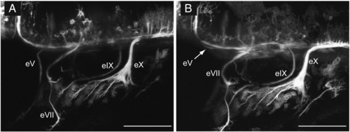
Only bmnV efferent pathfinding is affected in ngn1 morphants. Panel A is a lateral view of a control nkx:mgfp embryo at 52 hpf showing the typical appearance of branchiomeric efferent axons. Panel B is a lateral view of an embryo injected with the ngn1-MO at 52 hpf. As can be seen, only the bmnV efferents (indicated by the arrow) exhibited abnormal pathfinding as they traveled in an anterior, rather than ventral, direction: the other three branchiomeric efferents project normally. eV, bmnV efferents; eVII, bmnVII efferents; eIX, bmnIX efferents; eX, bmnX efferents. In both images, anterior is to the left and dorsal at the top. Scale bars are 100 μm. |
|
Gli1 null mutants exhibit defects in the central afferent projections of gIX and gX, but not gVII. A: lateral view of a control 4 dpf 3.2:gfp larva; B and C: lateral view of GFP+ neurons and their axons in two different 4 dpf gli1 - / -;3.2:gfp null mutants. Notice misrouting of the aIX and aX axons in B and aX in C. In both larvae aVII is unaffected. aVII, central afferents of gVII; aIX, central afferents of gIX; aX, central afferents of gX; dotted circle indicates the otic vesicle. Anterior to left, dorsal at top. Scale bars are 100 μm. |
|
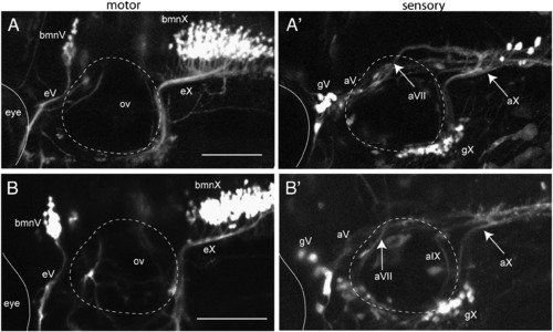
Hh signaling does not have a direct role on central sensory axon pathfinding. Images are from 4 dpf larvae co-expressing isl:gfp (left panels) and 3.2.cherry (right panels). The images shown are representative of 4 control and 7 treated embryos imaged from 2 separate experiments. Panels A, A′ is a control larva (0.5% ethanol); B,B′ cycA treatment (50 μM cycA/0.5% ethanol) beginning at 24 hpf shows no effect on either sets of axons. Dotted circle is otic vesicle (ov), eyes are demarcated by thin solid line. Arrows in A′ and B′ point to vagal afferents entering hindbrain. bmnV, motor nucleus of V; bmnX, vagal motor nucleus; eV, efferent nerve of bmnV; eX, efferent nerve of bmnX; aV, afferent nerve of gV; gV, trigeminal sensory ganglion; gX, vagal sensory ganglion; aVII, afferent nerve of gVII; aIX, afferent nerve of gIX; aX, afferent nerve of gX. Anterior to left, dorsal at top. Scale bars are 100 μm. EXPRESSION / LABELING:
|
|
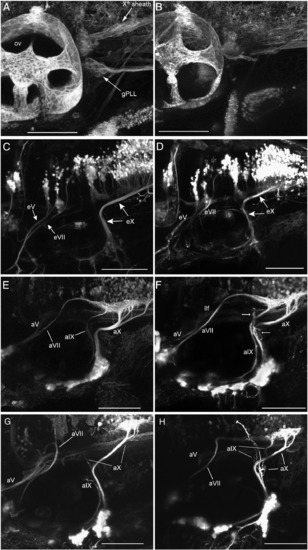
foxd3 nulls have lost peripheral glia, resulting in some cranial afferents becoming defasciculated and misrouted. A: 4 dpf control foxd3+/ -sox:rfp larva, B: 4 dpf mutant foxd3 - / -;sox:rfp larva showing loss of the Xth sheath and gPLL satellite glia. C: 4 dpf control foxd3+/ -;isl:gfp larva, D: 4 dpf mutant foxd3 - / -;isl:gfp larva showing correct proximal eV, eVII and eX projections. E: 4 dpf control foxd3+/ -3.2:gfp larva, F–H: 4 dpf mutant foxd3 - / -3.2:gfp larvae show a spectrum of axon misrouting; subsets of aVII (G), aIX (G,H) and aX (F–H) afferent axons are defasciculated and misrouted (e.g., arrows in F), whereas the remaining axons project correctly. gPLL, posterior lateral line ganglion satellite cells; ov, otic vesicle; eV, efferent nerve of bmnV; eVII, efferent nerve of bmnVII; eX, efferent nerve of bmnX; aV, afferent nerve of gV; aVII, afferent nerve of gVII; aIX, afferent nerve of gIX; aX, afferent nerve of gX; llf, lateral longitudinal fascicle. Anterior to left, dorsal at top. All scale bars are 100 μm. EXPRESSION / LABELING:
PHENOTYPE:
|
|
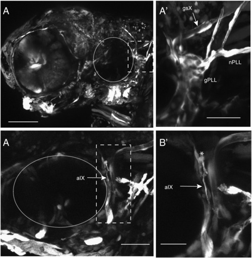
Expression of Foxd3 in cranial sensory neurons does not rescue afferent misrouting in foxd3 null mutants. Panel A, lateral view of 4 dpf mutant foxd3 - / - larva exhibiting a mosaic pattern of Foxd3/GFP co-expressing cells. A′ is higher power image of the boxed region in A and shows that expression of Foxd3 can rescue cranial peripheral glia, as shown by the GFP+ sheaths surrounding the Xth and lateral line nerves (nPLL). In addition, satellite glia are seen in sensory ganglia (gPLL). B: In a different rescued larva, expression of Foxd3 in gIX neurons did not prevent defasciculation of their afferent axons: a higher power image of the boxed region is shown in B′. An asterisk shows misrouted aIX axons that are not sheathed by GFP+ cells. Dotted circle, otic vesicle; gsX, glial sheath of afferent X; gPLL, posterior lateral line ganglion satellite glia; nPLL, posterior lateral line nerve glial cells; aIX, afferent axons of gIX. Anterior to left, dorsal at top. Scale bars: A, 100 μm; A′, 50 μm; B, 50 μm; B′, 25 μm. |
Reprinted from Developmental Biology, 357(2), Cox, J.A., Lamora, A., Johnson, S.L., and Voigt, M.M., Diverse mechanisms for assembly of branchiomeric nerves, 305-17, Copyright (2011) with permission from Elsevier. Full text @ Dev. Biol.

