
Fig. 2
- ID
- ZDB-IMAGE-111019-2
- Genes
- Publication
- Cox et al., 2011 - Diverse mechanisms for assembly of branchiomeric nerves
- All Figures
- Figures for Cox et al., 2011
|
Fig. 2
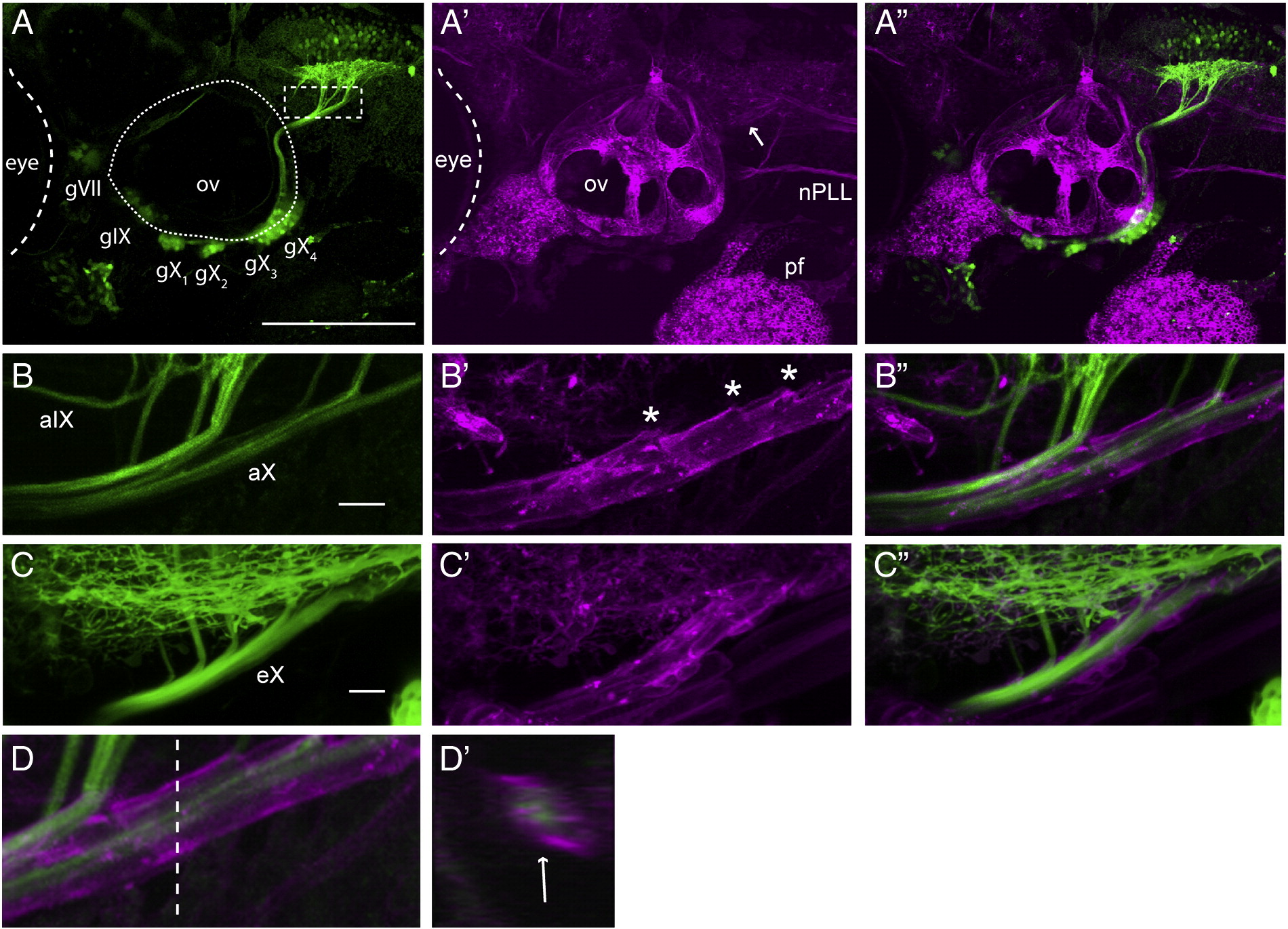
Cranial nerves containing sensory and motor axons are ensheathed by glia by 4 dpf. A shows a representative lateral view of GFP-expressing afferent axons in a 3.2:gfp;sox:rfp larva (n = 17), A′ shows RFP-expressing cells in the same larva (the arrow indicates the position of the vagal nerve sheath), and A" is a merge of A and A′. B is a higher power of the area indicted by the dashed rectangle in A showing GFP+ vagal afferents entering the hindbrain in a 3.2:gfp;sox:rfp larva; B′ shows the RFP+ glial sheath; B" is a merge of B and B′. Asterisks mark openings in the glial sheath where the vagal afferents leave the sheath to enter the hindbrain proper. C shows a lateral view of the bmnX efferents exiting the hindbrain in a 4 dpf nkx:mgfp;sox:rfp larva (n = 5) (also seen in green are oligodendrocyte processes in the hindbrain); C′ shows the glial sheath and C" is a merge of C and C′. D is a lateral view of aX in a 3.2:gfp;sox:rfp larva, D′ is an orthogonal projection (cross-section), taken at the level of the dotted line in D, showing the ensheathment of the peripheral portion of aX (indicated by arrow). gVII–gX, sensory ganglia; ov, otic vesicle; nPLL posterior lateral line nerve; aIX and aX- afferent nerves of gIX and gX; eX- efferent nerve of bmnX, pf, pectoral fin. All images are lateral, with anterior to left, dorsal at top. Scale bars: A: 100 μm, B: 20 μm, C: 20 μm.
Reprinted from Developmental Biology, 357(2), Cox, J.A., Lamora, A., Johnson, S.L., and Voigt, M.M., Diverse mechanisms for assembly of branchiomeric nerves, 305-17, Copyright (2011) with permission from Elsevier. Full text @ Dev. Biol.