
- Title
-
Aberrant expression of genes necessary for neuronal development and notch signaling in an epileptic mind bomb zebrafish
- Authors
- Hortopan, G.A., and Baraban, S.C.
- Source
- Full text @ Dev. Dyn.
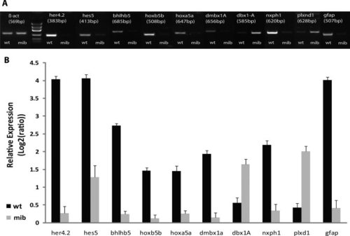
A,B: Gene expression detection of all the genes of interest (gfap included), using (A) reverse transcriptase-polymerase chain reaction (RT-PCR) in 2% ethidium bromide agarose gel electrophoresis (A) and quantitative real-time PCR (qPCR; B). Levels of mRNA, measured by qPCR were normalized to β-act. Error bars indicate ± SEM. Student′s t-test, P < 0.05. bhlhb5, basic helix-loop-helix domain containing, class B, 5; dbx1a, developing brain homeobox 1a; dmbx1a, diencephalon/mesencephalon homeobox 1a; gfap, glial fibrillary acidic protein; her4.2, hairy-related 4.2; hes5, hairy and enhancer of split 5; hoxa5a, homeo box A5a; hoxb5b, homeobox protein (hoxb5b) gene; nxph1, neurexophilin 1; plxnd1, plexin D1. EXPRESSION / LABELING:
|

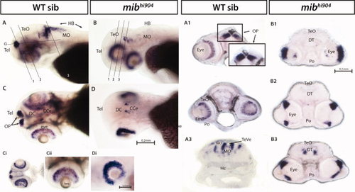
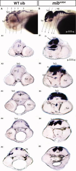
her4.2 mRNA expression in wild-type (WT) sibling (left panels) and mibhi904 mutant zebrafish (right panels) by in situ hybridization. A,B: Whole-mounts are shown in lateral views. A1–A5: WISH transversal cryostat sections of a stage 72 hr postfertilization (hpf) WT sibling at levels illustrated by the dashed lines in A. B1–B5: WISH transversal cryostat sections of a stage 72 hpf mibhi904 mutant at levels illustrated by the dashed lines in B. Note that dashed lines were indicatively drawn here (and in all subsequent figures) to reflect the approximate cutting angle and location. ac, anterior commissure; DC, diencephalon; DT, dorsal thalamus; EmT, eminentia thalami; H, hypothalamus; HB, hindbrain; Hc, caudal hypothalamus; Hi, intermediate hypothalamus; lfb, lateral forebrain bundle; MO, medulla oblongata; P, pallium; Po, preoptic region; Poc, postoptic commissure; Pr, pretectum; PTd, dorsal part of posterior tuberculum; PTv, ventral part of posterior tuberculum; S, subpallium; T, midbrain tegmentum; Tel, telencephalon; TeO, tectum opticum; VT, ventral thalamus. Scale bar = 0.2 mm in A,B, 0.1 mm in A1–A5,B1–B5. EXPRESSION / LABELING:
|

hes5 mRNA. A,B: Whole-mounts are shown in lateral views. A1–A4: Whole-mount in situ hybridization (WISH) transversal cryostat sections of a stage 72 hpf WT. B1–B4: WISH transversal cryostat sections of a stage 72 hpf mibhi904. Note that it is difficult to precisely match thin cut sections of the larval zebrafish (e.g., eye is smaller in B3 and B4) and this does not reflect an asymmetrical expression pattern in the midbrain. Ha, habenula; Hr, rostral hypothalamus; m, medial tectum opticum; PTv, ventral part of posterior tuberculum; TeVe, tectal ventricle; TS, torus semicircularis; TVe, telencephalic ventricle. Scale bar = 0.2 mm in A,B, 0.1 mm in A1–A4,B1–B4. EXPRESSION / LABELING:
|
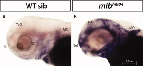
bhlhb5 mRNA expression. A–D: Whole-mounts are shown in lateral (A,B) and dorsal views (C,D). Ci,Di: Smaller panels show a higher magnification of the eye, in lateral views. Note the mibhi904 mutant eye where the retinal layers fail to differentiate. A1–A3: Whole-mount in situ hybridization (WISH) transversal cryostat sections of a stage 72 hours postfertilization (hpf) wild-type (WT). A4–A5: WISH coronal cryostat sections of a stage 72 hpf WT. B1–B3: WISH transversal cryostat sections of a stage 72 hpf mibhi904 mutant. Abbreviations as above: CCe, cerebellum; INL, inner nuclear layer; GCL, ganglion cell layer; ONL, outer nuclear Layer; OP, olfactory pits. Scale bar = 0.2 mm in A–D, 0.1 mm in A1–A5,B1–B3, 0.05 mm in Ci,Di.Figure 5. The hoxa5a and hoxb5b mRNA expression. A,C,E,G: In the upper panels, whole-mounts are shown in lateral views. B,D,F,H: Lower panels are higher magnification and same orientation of area framed in A, C, E, G. Abbreviations as above: Hr, hindbrain rhombomeres; PA, pharyngeal arch. Scale bars = 0.2 mm in A,C,E,G, 0.1 mm in B,D,F,H. EXPRESSION / LABELING:
PHENOTYPE:
|
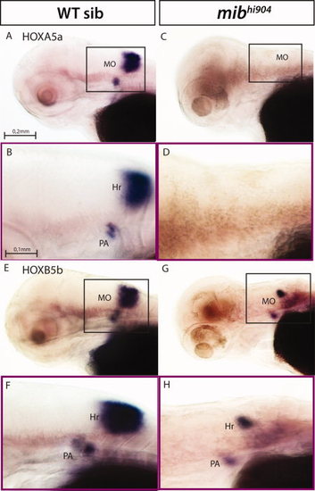
The hoxa5a and hoxb5b mRNA expression. A,C,E,G: In the upper panels, whole-mounts are shown in lateral views. B,D,F,H: Lower panels are higher magnification and same orientation of area framed in A, C, E, G. Abbreviations as above: Hr, hindbrain rhombomeres; PA, pharyngeal arch. Scale bars = 0.2 mm in A,C,E,G, 0.1 mm in B,D,F,H. EXPRESSION / LABELING:
|
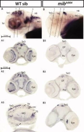
The dmbx1a mRNA expression. A,B: Whole-mounts are shown in lateral views. A1–A3: Whole-mount in situ hybridization (WISH) transversal cryostat sections of a stage 72 hours postfertilization (hpf) wild-type (WT). B1–B3: WISH transversal cryostat sections of a stage 72 hpf mibhi904. Abbreviations (as above): cec, cerebellar commissure; Ch, chorda dorsalis; Poc, postoptic commissure; Va, valvula cerebelli. Scale bars = 0.2 mm in A,B, 0.1 mm in A1–A4,B1–B3. EXPRESSION / LABELING:
|
dbx1a mRNA expression. A,B: Whole-mounts are shown in lateral views. A1–A5: Whole-mount in situ hybridization (WISH) transversal cryostat sections of a stage 72 hours postfertilization (hpf) wild-type (WT). B1–B5: WISH transversal cryostat sections of a stage 72 hpf mibhi904. Abbreviations as above. Scale bars = 0.2 mm in A,B, 0.1 mm in A1–A5,B1–B5. EXPRESSION / LABELING:
|
plxnd1 mRNA. A,B: Whole-mounts are shown in lateral views. Abbreviations as above. Scale bars = 0.2 mm in A,B. EXPRESSION / LABELING:
|
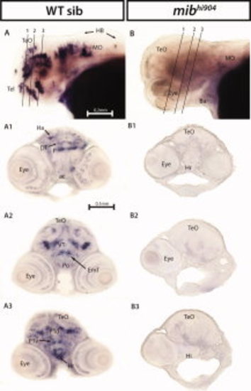
nxph1 mRNA. A,B: Whole-mounts are shown in lateral views. A1–A3: Whole-mount in situ hybridization (WISH) transversal cryostat sections of a stage 72 hours postfertilization (hpf) wild-type (WT). B1–B3: WISH transversal cryostat sections of a stage 72 hpf mibhi904. Abbreviations as above: PTd, dorsal part of posterior tuberculum; PTv, ventral part of posterior tuberculum. Scale bars = 0.2 mm in A,B, 0.1 mm in A1–A3,B1–B3. EXPRESSION / LABELING:
|
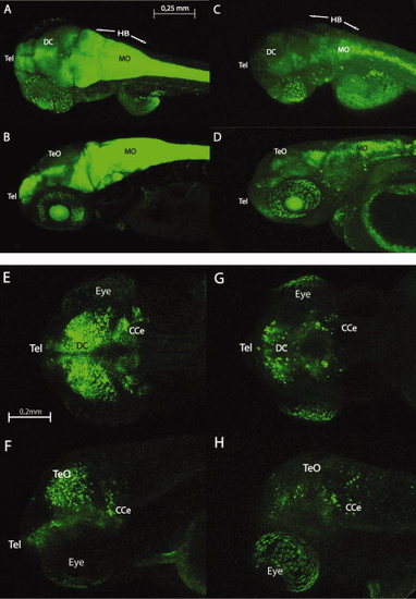
A–H: Confocal live whole-mount images of Gfap:GFP (upper panel) and Dlx5-6:GFP transgenic zebrafish brain (lower panel) in wild-type (left side) and mibhi904 mutant (right side) at 3 days postfertilization (dpf): dorsal views (A,C,E,G) and lateral views (B,D,F,H). The transgenic line for the glial fibrillary acidic protein (GFAP) drove expression (shown as green fluorescence) initially in the telencephalon and then in the eye and midbrain. Gfap:GFP is also highly prominent in hindbrain in wild-type (WT)siblings with a weak expression pattern in mibhi904 mutants. Dlx5-6:GFP in mibhi904 mutants is reduced with only few cells showing expression in diencephalon, eyes, and in the cerebellar region. Images of live zebrafish were obtained using a Leica SP5 confocal scanning fluorescence microscope. Abbreviations as above. Scale bars = 0.25 mm in A–D, and 0.2 mm in E–H. EXPRESSION / LABELING:
|