
- Title
-
Characterization and comparative expression of zebrafish calpain system genes during early development
- Authors
- Lepage, S.E., and Bruce, A.E.
- Source
- Full text @ Dev. Dyn.
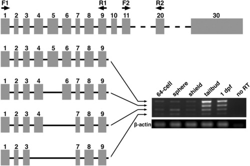
Temporal expression pattern and exon organization of four calpastatin splice variants. Semiquantitative reverse transcriptase (RT) -polymerase chain reaction using cast F1 and R1 primers was used to determine the relative abundance of four cast transcripts at 64-cell, sphere, shield, bud, and 1 day postfertilization (dpf). One representative no template control (no RT) is shown and β-actin was used as a loading control. Schematic representation of exon organization is shown for each of the four transcripts. cast F1 and R1 primers amplify exons 1-9 and F2 and R2 amplify exons 11-20 as indicated. cast F2 and R2 amplify a single product (see Fig. 3). |
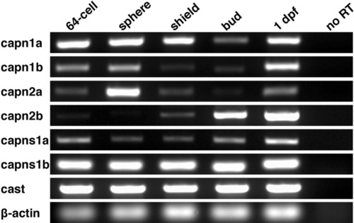
Semiquantitative reverse transcriptase (RT) -polymerase chain reaction showing the temporal expression patterns of zebrafish CAPN1 (capn1a, 1b), CAPN2 (capn2a, 2b), CAPNS1 (capns1a, 1b), and CAST (cast) orthologs. The relative abundance of transcript at 64-cell, sphere, shield, bud, and 1 day postfertilization (dpf) was determined for each ortholog. A representative no template control (no RT) is shown for each primer set and β-actin was used as a loading control. |
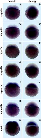
Whole-mount in situ hybridization analysis of zebrafish CAPN1, CAPN2, CAPNS1, and CAST orthologs during cleavage and blastula stages. A-N: Spatial expression of capn1a (A,B), capn1b (C,D), capn2a (E,F), capn2b (G,H), capns1a (I,J), capns1b (K,L), and cast (M,N) is shown for representative cleavage stage embryos at the 4-cell (A,C,E,G,I,K,M) and blastula stage embryos at the oblong stage (B,D,F,H,J,L,N). All embryos are shown in lateral view. |

Spatial expression patterns of zebrafish capn1, capn2, capns1, and cast genes during gastrulation, segmentation, and pharyngula stages. A-ZZZ: Representative whole-mount in situ hybridizations showing distribution of capn1a (A-D), capn1b (E-H), capn2a (I-L), capn2b (M-P), capns1a (Q-T), capns1b (U-X), and cast (Y-ZZZ) transcripts at shield stage (A,E,I,M,Q,U,Y), four to six somites (B,F,J,N,R,V,Z), 1 day (C,G,K,O,S,W,ZZ), and head regions of 2 day embryos (D,H,L,P,T,X,ZZZ). All embryos are shown in lateral view. Shield and 4-6 somite stage embryos are oriented with dorsal to the right. One day postfertilization (dpf) and 2 dpf embryos are oriented with anterior to the left. At 2 dpf, no staining was observed in structures posterior to the fin bud. Arrowheads indicate anterior mesendoderm (F,J,Z), polster (N,R), and proctodeum (O,S,W). Arrows indicate pronephros (S,W), pharynx (P,T,X,ZZZ), and hindbrain neuron clusters (H). The asterisk in W indicates the mid-hindbrain boundary region. hg, hatching gland. |
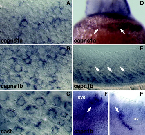
Tissue-specific expression of capns1a, capns1b, capn1b, and cast by whole-mount in situ hybridization. A-C: Higher magnification views of the enveloping layer (EVL) expression of capns1a (A), capns1b (B), and cast (C) in flat-mounted 4-6 somite stage embryos shown in Figure 5R,V,Z. D: Ventral view of 1 day postfertilization (dpf) embryo shown in Figure 5S, showing expression of capns1a in the hatching gland (arrows). E-F′: Higher magnification views of capn1b expression in medial somites at 1 dpf (E, arrows) and two groups of bilateral (left side shown) hindbrain neuron clusters at 2 dpf (F,F′, arrows). Embryo in E is shown in lateral view and embryos in F and F′ are viewed dorsally. All embryos E-F′ are oriented with anterior to the left. Position of the eye and otic vesicle (ov) are indicated in F and F′, respectively. EXPRESSION / LABELING:
|