
Fig. 5
- ID
- ZDB-IMAGE-080402-44
- Genes
- Publication
- Lepage et al., 2008 - Characterization and comparative expression of zebrafish calpain system genes during early development
- All Figures
- Figures for Lepage et al., 2008
|
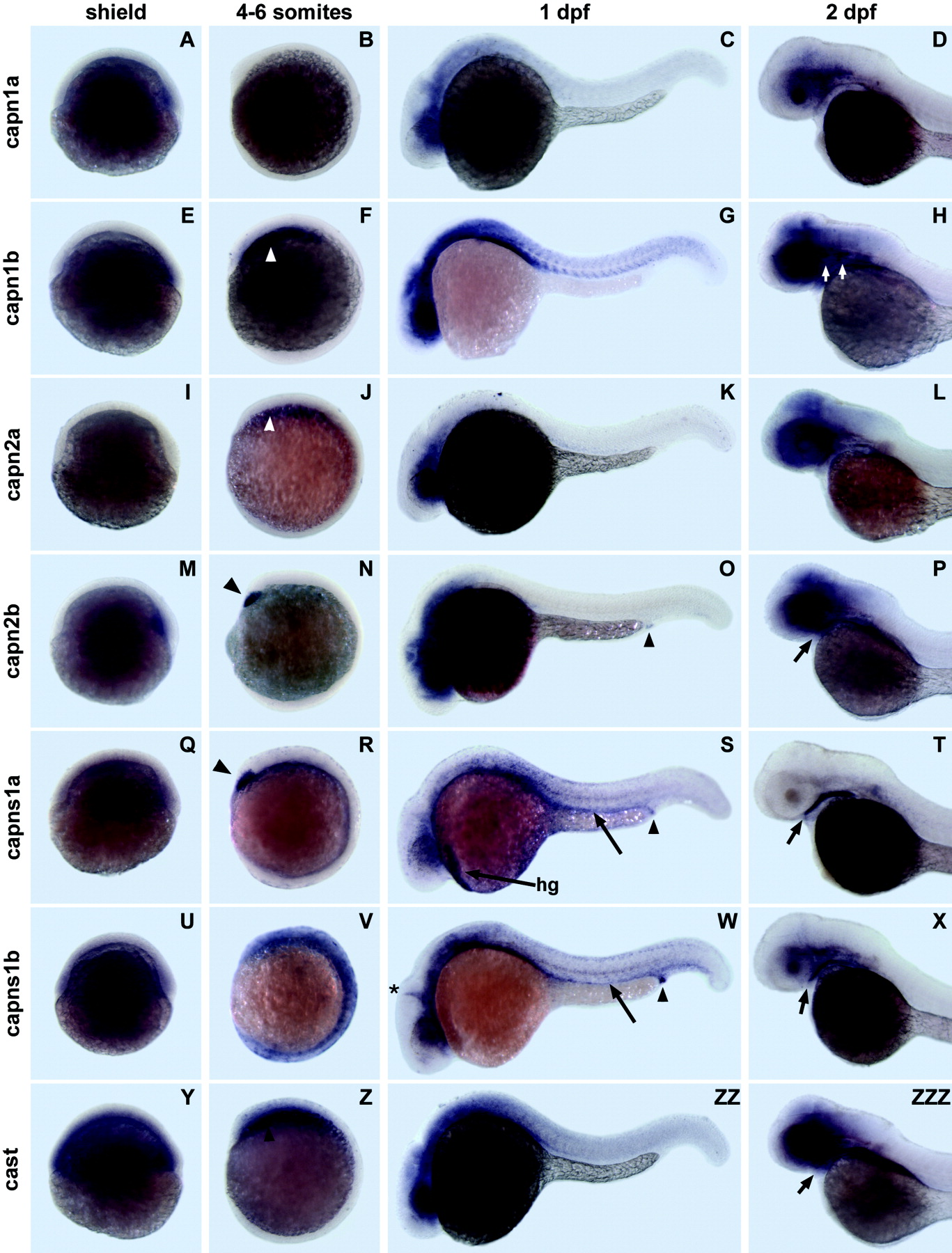
Fig. 5 Spatial expression patterns of zebrafish capn1, capn2, capns1, and cast genes during gastrulation, segmentation, and pharyngula stages. A-ZZZ: Representative whole-mount in situ hybridizations showing distribution of capn1a (A-D), capn1b (E-H), capn2a (I-L), capn2b (M-P), capns1a (Q-T), capns1b (U-X), and cast (Y-ZZZ) transcripts at shield stage (A,E,I,M,Q,U,Y), four to six somites (B,F,J,N,R,V,Z), 1 day (C,G,K,O,S,W,ZZ), and head regions of 2 day embryos (D,H,L,P,T,X,ZZZ). All embryos are shown in lateral view. Shield and 4-6 somite stage embryos are oriented with dorsal to the right. One day postfertilization (dpf) and 2 dpf embryos are oriented with anterior to the left. At 2 dpf, no staining was observed in structures posterior to the fin bud. Arrowheads indicate anterior mesendoderm (F,J,Z), polster (N,R), and proctodeum (O,S,W). Arrows indicate pronephros (S,W), pharynx (P,T,X,ZZZ), and hindbrain neuron clusters (H). The asterisk in W indicates the mid-hindbrain boundary region. hg, hatching gland.