|
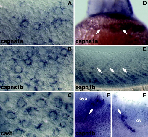
Tissue-specific expression of capns1a, capns1b, capn1b, and cast by whole-mount in situ hybridization. A-C: Higher magnification views of the enveloping layer (EVL) expression of capns1a (A), capns1b (B), and cast (C) in flat-mounted 4-6 somite stage embryos shown in Figure 5R,V,Z. D: Ventral view of 1 day postfertilization (dpf) embryo shown in Figure 5S, showing expression of capns1a in the hatching gland (arrows). E-F′: Higher magnification views of capn1b expression in medial somites at 1 dpf (E, arrows) and two groups of bilateral (left side shown) hindbrain neuron clusters at 2 dpf (F,F′, arrows). Embryo in E is shown in lateral view and embryos in F and F′ are viewed dorsally. All embryos E-F′ are oriented with anterior to the left. Position of the eye and otic vesicle (ov) are indicated in F and F′, respectively.
|