- Title
-
An olig2 reporter gene marks oligodendrocyte precursors in the postembryonic spinal cord of zebrafish
- Authors
- Park, H.C., Shin, J., Roberts, R.K., and Appel, B.
- Source
- Full text @ Dev. Dyn.
|
Transverse sections of spinal cords of Tg(olig2:egfp) transgenic zebrafish, dorsal to top. Strong EGFP fluorescence was evident in ventral cells with radial processes extending to the pial surface at all stages (arrows). A: At 3 dpf, other EGFP+ cells had multiple fine membrane processes and occupied positions characteristic of OPCs (arrowheads). B-D: The number of EGFP+ radial cells in transverse sections remained constant at all stages examined through 3 months whereas the number of non-radial EGFP+ cells increased throughout the spinal cord. Scale bar = (A) 20 μM; (B) 30 μM; (C) 50 μM; (D) 80 μM. |
|
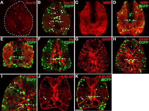
olig2+ cells include radial glia and oligodendrocyte lineage cells in the postembryonic spinal cord. All images are single confocal optical sections of transverse sections of 30-dpf Tg(olig2:egfp) zebrafish spinal cords, dorsal up. Arrows indicate the soma and processes of EGFP+ radial cells. A,B: Anti-Sox10 labeling (A) and combined anti-Sox10 and EGFP images of same section (B). Most EGFP+ cells were Sox10+ oligodendrocyte lineage cells (arrowheads) except for EGFP+ radial glia. C,D: Anti-MBP antibody labeling (C) and combined MBP and EGFP images of same section (D). E,F: Laterally located EGFP+ cells were dispersed throughout large tracts of anti-acetylated Tubulin-positive axonal fibers (E), but there was no co-localization of EGFP and Hu (F). G-I: Anti-GFAP antibody labeling (G) and combined images of the same section for GFAP and EGFP (H, I). EGFP+ cell bodies were closely associated with GFAP+ processes but fluorescent signals for EGFP and GFAP did not appear to colocalize. J,K: Anti-BLBP antibody labeling (J) and combined images of the same section for BLBP and EGFP (K). Both the soma and processes of EGFP+ radial cells were BLBP+. Scale bar = 50 μM for all panels except I, for which it represents 100 μM. EXPRESSION / LABELING:
|
|
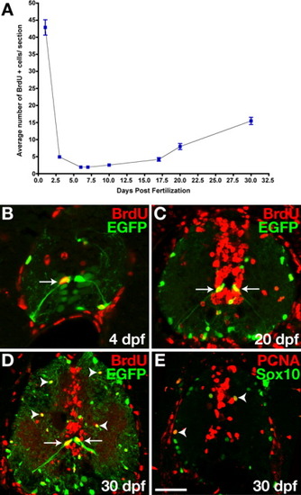
olig2+ spinal cord cells divide at postembryonic stages. All images are single confocal optical sections of transverse sections of 30 dpf Tg(olig2:egfp) zebrafish spinal cords, dorsal up. A: Graph showing average number of BrdU+ spinal cord cells per transverse section at different embryonic and postembryonic stages. Data were compiled from 15-30 sections per timepoint. Bars represent standard error of the mean. B: 4 dpf Tg(olig2:egfp) transgenic larvae pulse-treated with BrdU and labeled with anti-BrdU antibody. Arrows indicate EGFP+ radial glia, which incorporated BrdU. C,D: Twenty- and 30-dpf Tg(olig2:egfp) transgenic fish treated with BrdU repeatedly. Many cells, including EGFP+ radial glia (arrows), bordering the central canal and medial septum incorporated BrdU. Some EGFP+ oligodendrocyte lineage cells were also BrdU+ (arrowheads). E: Double labeling with anti-Sox10 and anti-PCNA antibodies. Sox10+ PCNA+ cells were proliferative OPCs (arrowheads). Scale bar = 30 μM (B,C), 50 μM (D,E). EXPRESSION / LABELING:
|
|
olig2+ radial glial cells have asymmetric features. A-C: Transverse sections of 15-dpf Tg(olig2:egfp) fish, dorsal up. A: aPKC (red) was localized to the apical membranes of EGFP+ radial glia (green) at the central canal. B: Two BrdU+ EGFP+ cells (arrows) were next to each other near the central canal. These cells might have been recently divided siblings. C: Anti-phospho-Histone H3 (pH3) antibody labeling revealed chromosomes (arrows) aligned with the axis of division perpendicular to the central canal (dashed line). Scale bar = 10 μM. EXPRESSION / LABELING:
|


