
- Title
-
SMC3 knockdown triggers genomic instability and p53-dependent apoptosis in human and zebrafish cells
- Authors
- Ghiselli, G.
- Source
- Full text @ Mol. Cancer

Expression of zebrafish smc3 during embryogenesis. A) Semiquantitative RT-PCR analysis of smc3 transcript level in zebrafish embryos at different times during development. ef-1 transcript level, which is expressed throughout development, was used as internal control. B) Expression of constituents of the cohesin multimeric protein complex: zebrafish smc1, rad21 and sa2 transcripts were quantitated as described in panel A. C) Spatio-temporal expression of smc3 mRNA. Whole-mount mRNA in-situ hybridization was performed at the indicated developmental stages. Left-to-right: lateral view with the animal pole up of embryos at 50% epiboly (6 hpf), at 90% epiboly (9 hpf), at 3-somite stage (12 hpf), and at 24 hpf. EXPRESSION / LABELING:
|

Effect of Smc3 knockdown on embryogenesis. A) Western immunoblot analysis of Smc3 level in embryos injected at 1–2 cell stage with increasing doses of smc3-MO or smc3-mmMO. At 24 hpf 10 embryos were dechorionated and the yolk remnant removed. After homogenization in 100 μl lysis buffer, proteins were separated on 7.5% SDS-PAGE and Smc3 detected by immunoblotting using goat anti-human SMC3 antibody followed by ECL reaction. B) Morphogenesis in normal (a-e) and morphant (f-j) embryos injected with 8 ng of smc3-MO. At the indicated times embryos were collected and their morphology examined. Note the presence of necrotic cells in the eye and the cerebellar region of embryos at 15 hpf. At 24 hpf the majority of the morphants display head necrosis and a range of tail developmental defects including a reduction of the tail extension, the loss of chevron-like structure of the somites and the narrowing or disappearance of the notocord. EXPRESSION / LABELING:
PHENOTYPE:
|
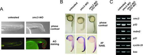
p53 pathway activation and cell death following Smc3 knockdown. A) Acrydine Orange (AO) staining of apoptotic cells. Embryos at 1-2-two cell stage were injected with 1 nl of Danieau buffer (untreated group) or with 8 ng/embryo smc3-MO and at 24 hpf incubated in E3 medium containing 5 μg/ml of the fluorescent dye. Following 1 h incubation, the embryos were washed in PBS and observed under a fluorescence microscope. Note the high level of fluorescence signal at the terminal edge of the morphant's tail whereas in the untreated embryo fluorescence is only detected at the prospective vent. B) Morphological assessment and TUNEL immunostaining of apoptotic and dead cells in morphants. Embryos were injected at 1–2 cell-stage with MO to block translation of either smc3 only or of smc3 and p53, and examined at 28 hpf. C) Semi-quantitative RT-PCR of smc3, p53 and a p53-target genes transcript level in untreated embryos and in morphants at 24 hpf |
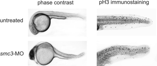
Effect of Smc3 knockdown on the phosphorylation in vivo of histone H3. Embryos at 1-2-two cell stage were injected with the vehicle alone or with 8 ng/embryo smc3-MO and analyzed at 24 hpf. Embryos were fixed overnight in 4% para-formaldehyde and permeabilized by immersion in -20°C acetone for 10 min. After overnight incubation at 4°C with 1 μg/ml rabbit anti-pH3 antibody, the embryos were placed in 1 μg/ml HRP-conjugated goat anti-rabbit IgG solution and the immunocomplexes detected by reaction with DAB reagent. The punctuate staining pattern visible in the embryo tails identifies the localization of the pH3 antigen and thus of the mitotic cells. |
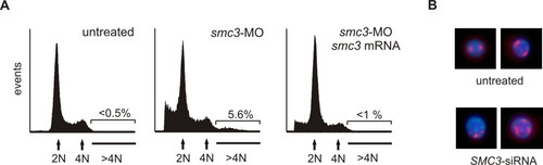
Cell DNA content analysis and chromosomal abnormalities following SMC3 knockdown. A) Cell cycle FACS analysis of zebrafish embryonic cells. Embryos at 1–2 cell stage were injected with 1 nl Danieau buffer (left-hand side) or 4 ng of smc3-MO (center) or 4 ng of smc3-MO together with 250 pg smc3 mRNA (right-hand side). Batches of 30 embryos at 24 hpf were incubated in 0.05% trypsin 15 min at 25°C and the cells dissociated by pipetting through a large bore glass pipette. After fixation in 70% ethanol at 4°C overnight, cells were stained with propidium iodine/RNase, and the DNA cell content analyzed by flow cytometry. Gated data were used for analysis. Note the presence of a population of aneuploid (DNA > 4) cells in embryos following Smc3 knockdown. B) Analysis of the centrosome organization in human cells. Human 293 cells were transfected with 50 ng/ml of SMC3-siRNA and after 24 h plated on a poly-lysine-coated coverslip. Fourty-eight h later the cells were fixed in 4% para-formaldehyde, permebilized and then incubated with 1:1000 anti-human γ-tubulin antibody. Following incubation with Alexafluor 567-conjugated anti-rabbit IgG (Molecular Probes) and DNA staining with DAPI, the cells were examined under a fluorescence/UV microscope and the red (Alexafluor 567) and blue (DAPI) signals combined. PHENOTYPE:
|

Unillustrated author statements PHENOTYPE:
|