- Title
-
Zebrafish protocadherin 10 is involved in paraxial mesoderm development and somitogenesis
- Authors
- Murakami, T., Hijikata, T., Matsukawa, M., Ishikawa, H., and Yorifuji, H.
- Source
- Full text @ Dev. Dyn.
|
Ectopic expression of tagged Pcdh10 in zebrafish embryos. Single blastomeres of eight-cell embryos were injected with an RNA of GFP-tagged Pcdh10 construct, Pcdh10G. a: Confocal microscopy of 11 hours postfertilization (hpf) embryos expressing Pcdh10G revealed that fluorescent cells remained coherent during gastrulation to form patches (arrowheads) on the embryo. In this particular embryo, the left segmental plate was split from the notochord (arrow) at such a patch of green cells. GFP image (green) was superimposed on a phalloidin image (red). Left-side oblique view with rostral to the top. b: Confocal image of a 11 hpf embryo injected with GFP control RNA showed green cells dispersed over the embryo. Left-side view with rostral to the top. c: Detailed confocal scan of a 9 hpf embryo showing Pcdh10G localized to the cell peripheries (green, arrowheads). Red, phalloidin; blue, 4,6-diamidino-2-phenylindole (DAPI). The animal pole is to the top, dorsal to the right. a and b, Z projections of serial optical sections. c, a single section. Scale bars = 50 μ m. |
|
Expression of pcdh10 in zebrafish embryos. Zebrafish embryos were stained by whole-mount in situ hybridization for pcdh10 or myoD. a-f: myoD. g-r: pcdh10. a-l: Dorsal views of the developing somites. m-r: Lateral views. a,g,m, 10 hours postfertilization (hpf); b,h,n, 11.5 hpf; c,i,o, 14 hpf; d,j,p, 16 hpf; e,k,q, 19 hpf; f,l,r, 22 hpf. Closed arrowheads indicate the latest visually segmenting somites. Closed arrows, epiphysis. Open arrowheads, otic vesicles. Open arrows, head mesoderm. Rostral is to the top. Scale bar = 100 μm. EXPRESSION / LABELING:
|
|
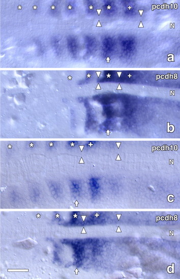
Comparison of expressions of pcdh10 and pcdh8 in paraxial mesoderm. Embryos were stained by whole-mount ISH for pcdh10 or pcdh8, manually sectioned, and flat-mounted for microscopy. a,b: 12 hours postfertilization (hpf). c,d: 14 hpf. a,c: pcdh10. b,d: pcdh8. Arrows indicate the latest visually segmenting somites. Asterisks indicate the older somites with positive pcdh10 or pcdh8 staining. Plus signs indicate the latest presumptive somites. Opposing arrowheads, adaxial cells; N, notochords. Dorsal views with the rostral to the left. Scale bar = 50 μm. EXPRESSION / LABELING:
|
|
Antisense morpholino and dominant-negative construct of Pcdh10 disturbed somitogenesis. b-f: Embryos were injected with pcdh10 antisense morpholino oligonucleotide (MO), pcdh10mo, fixed at 14-15 hours postfertilization (hpf), and stained by in situ hybridization (ISH) for MyoD to show somite phenotypes. Somitogenesis was scored as affected when (AS) left-right asymmetry was found in some somites (arrowheads in b); (BR) there were bridges (right arrowhead in c), compression of adjacent somites (arrowhead in d) or blurred somite boundaries (left arrowhead in c); or (MS) some somites were missing or misplaced (arrowheads in e), or the midline structures were widened and disorganized (f). a: Embryo injected with negative control MO with normal morphologies. g,h: Embryos were injected with Pcdh10ecF RNA at the four-cell stage. Embryos with unilateral expression of GFP as a tracking dye (arrowheads in g) were fixed for MyoD ISH. Note the deformation of somites (arrowheads in h) ipsilateral to GFP expression. Dorsal views with rostral to the top. Scale bars = 100 μm. EXPRESSION / LABELING:
|
|
pchd10 expression in the rostral structures. pcdh10 was expressed in the epiphysis (double arrowheads), diencephalon (arrowheads), myelencephalon (open arrows), otocyst (asterisk), eye muscles (closed arrow), and fin buds (closed arrows). a-c: Dorsal (a), lateral (b), and frontal (c) views of 33-hours postfertilization (hpf) embryos stained by whole-mount in situ hybridization for pcdh10. The dashed line in b indicates the focal plane of c. The focal plane of the inset in b is lateral to that outside the inset. Scale bar = 100 μm. EXPRESSION / LABELING:
|


