
IMAGE
Fig. 4
- ID
- ZDB-IMAGE-060203-2
- Genes
- Publication
- Murakami et al., 2006 - Zebrafish protocadherin 10 is involved in paraxial mesoderm development and somitogenesis
- All Figures
- Figures for Murakami et al., 2006
Image
|
Figure Caption
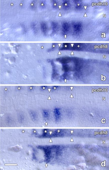
Fig. 4 Comparison of expressions of pcdh10 and pcdh8 in paraxial mesoderm. Embryos were stained by whole-mount ISH for pcdh10 or pcdh8, manually sectioned, and flat-mounted for microscopy. a,b: 12 hours postfertilization (hpf). c,d: 14 hpf. a,c: pcdh10. b,d: pcdh8. Arrows indicate the latest visually segmenting somites. Asterisks indicate the older somites with positive pcdh10 or pcdh8 staining. Plus signs indicate the latest presumptive somites. Opposing arrowheads, adaxial cells; N, notochords. Dorsal views with the rostral to the left. Scale bar = 50 μm.
Figure Data
Acknowledgments
This image is the copyrighted work of the attributed author or publisher, and
ZFIN has permission only to display this image to its users.
Additional permissions should be obtained from the applicable author or publisher of the image.
Full text @ Dev. Dyn.