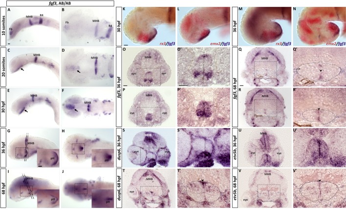
IMAGE

Fig. 1.

ID
ZDB-IMAGE-190723-2000
Genes
Source
Figures for Reuter et al., 2019
Image
Figure Caption

Fig. 1.

RNA in situ hybridisation reveals fgf3 expression in the developing hypothalamus. (A,B) At 10 somites, fgf3 transcripts are detectable in the forebrain (Fb), at the mid- hindbrain boundary (MHB) and in rhombomere 4 (R4). (C–F) At 20 somites and 30 hpf, fgf3 expression is present in the hypothalamic primordium (arrows), and (G–J) at 36 and 68 hpf in the posterior hypothalamus (pH). Left column depicts lateral views and right column ventral views. Insets are high magnifications of boxed areas. Anterior to the left. (K–N) Double in situ hybridisation for fgf3 with rx3 or emx2 at 30 and 36 hpf shows that fgf3 is concentrated to the posterior hypothalamus. Anterior to the left. (O–V) 20 µm frontal cryosections at levels indicated in G and I of embryos hybridised for fgf3, dusp6 and etv5b at 36 or 68 hpf. O′–V′ are high magnifications of boxed areas. Dashed lines in O′, P′, S′ and U′, and arrowheads in T′, Q′, R′ and V′ indicate ventricle. Dashed circles in T′, Q′, R′ and V′ outline borders of posterior hypothalamus. At 36 hpf, expression of fgf3 spans the entire medial to lateral dimension of the posterior hypothalamus. dusp6 and etv5b are similarly broadly expressed. At 68 hpf, the expression of fgf3 is restricted to cells located medially at the ventricle (arrowheads), while dusp6 and etv5b are present laterally, in cells around the posterior recess. Scale bars: 30 µm.

Figure Data
Acknowledgments
This image is the copyrighted work of the attributed author or publisher, and ZFIN has permission only to display this image to its users. Additional permissions should be obtained from the applicable author or publisher of the image. Full text @ Biol. Open