Fig. 3
Fig. 3
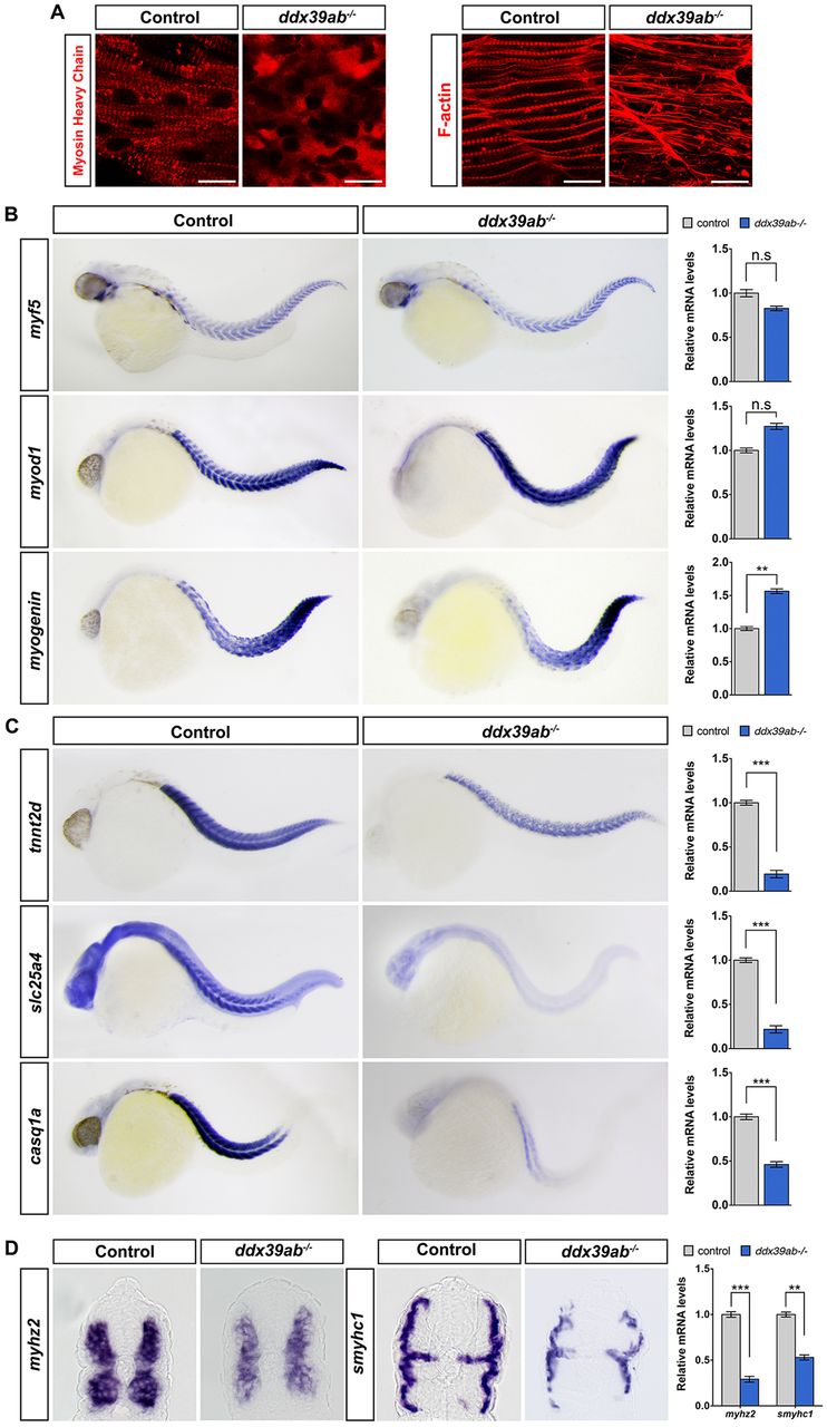
Loss of ddx39ab results in defective skeletal muscle differentiation in zebrafish. (A) Immunostaining demonstrates the organization of myofilaments in wild-type and ddx39ab mutant embryos at 24 hpf. Anti-MHC antibody (MF20) labels thick (myosin) filaments and F-actin was visualized by phalloidin staining. Scale bars: 20 μm. (B) RNA in situ hybridization for myogenic regulatory gene expression in wild-type and ddx39ab mutant zebrafish embryos at 32 hpf. (C,D) RNA in situ hybridization and qPCR analysis for myocyte structural gene expression in wild-type and ddx39ab mutant zebrafish embryos at 32 hpf. (B,C) Lateral views with anterior to the left. (D) Transverse section with dorsal side to top. At least 15 (A) or 20 (B-D) embryos of each genotype were analyzed and representative samples are shown. For qPCR results, data are mean±s.e.m. n.s, not significant. **P<0.01, ***P<0.001.

