
Fig. 6
- ID
- ZDB-IMAGE-160927-52
- Genes
- Publication
- Zhao et al., 2016 - Enhanced angiogenesis, hypoxia and neutrophil recruitment during Myc-induced liver tumorigenesis in zebrafish
- All Figures
- Figures for Zhao et al., 2016
|
Fig. 6
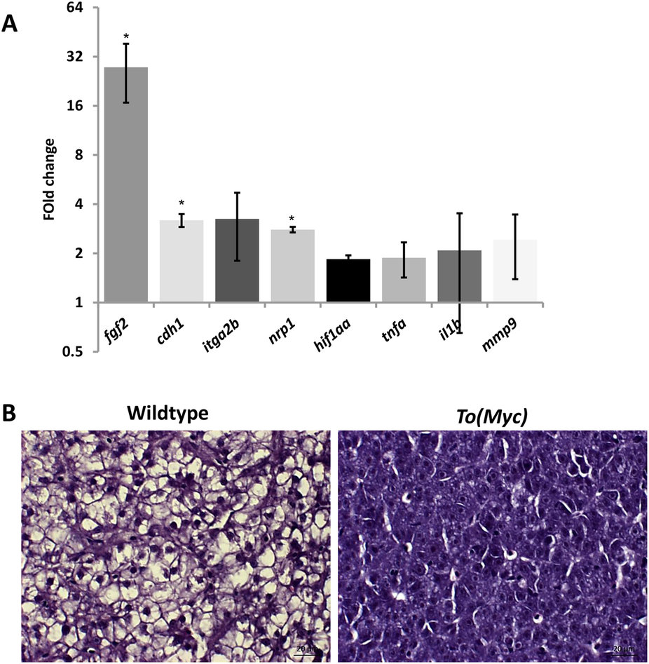
Molecular and histological characterization of Myc overexpressed livers.
One-month-old wildtype or TO(Myc) zebrafish were treated with 30 µg/ml Dox for 7 days and euthanized for RNA extraction and histological analyses. (A) Validation of increased angiogenesis, hypoxia and inflammatory response by biomarker gene expression. RNA expression of selected biomarker genes were measure by RT-qPCR. Fold changes shown are ratio of the values from To(Myc) fish over wildtype fish after calibration with beta-actin mRNA as an internal control. Asterisks indicate significant difference with P-value < 0.05 by t-test among the three biological replicates. (B) Histological comparison of livers from wildtype (left) and TO(Myc) (right) fish. Fish were treated with or without Dox (30 µg/ml) from 4 dpf to 7 dpf. Representative pictures are shown for each group (n = 10 per group). The original magnification was 100x. Scale bars = 20 µm.