
Fig. 4
- ID
- ZDB-IMAGE-150316-5
- Genes
- Publication
- Maier et al., 2014 - RA and FGF Signalling Are Required in the Zebrafish Otic Vesicle to Pattern and Maintain Ventral Otic Identities
- All Figures
- Figures for Maier et al., 2014
|
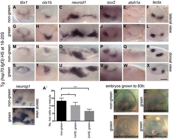
Fig. 4
Effects of fgf3 over-expression from the 18 somite stage on otic vesicle patterning.
Tg(hsp70:fgf3) embryos were heat shocked (HS) from 18S and sorted as non-green, non-transgenic siblings (A–F, M–R, Y, A′) or green transgenic embryos (G–L, S–X, Z, B′). (A–F, M–R, A′) In non-green embryos, expression of the non-neural markers tbx1 (n = 79; A,M) and otx1b (n = 52; B,N), the neuronal marker neurod1 (n = 18; C,O), and the sensory markers sox2 (n = 11), atoh1a (n = 40), tecta (n = 32) (D–F, P–R) and the neurogenic marker neurog1 (Y, n = 13) is normal. (G–L, S–X, B′) In Tg(hsp70:fgf3) embryos sorted as green, the expression of tbx1 is unaffected or slightly up-regulated (n = 97; G,S), while expression of otx1b (n = 56; H,T), neurog1 (n = 25; Z) and otic and non-otic neurod1 (n = 35; I,U) is strongly up-regulated. The expression of sox2 (n = 24), atoh1a (n = 83) and tecta (n = 100) is slightly reduced and the anterior part of the posterior domain marked by tecta is missing (J–L, V–X). (A′) Graphical representation of counts of GFP-positive sensory hair cells (pou4f3:GFP expression) in the demarcated area from embryos heterozygous for both Tg(hsp70:fgf3) and Tg(pou4f3:GFP). Error bars represent standard deviation. One-way ANOVA with Šídák′s multiple comparison post-test: *p<0.05, ***p<0.001. (B′–E′) Live images of Tg(hsp70:fgf3) embryos HS at 18S and grown to 83 hpf. The inner ear morphology of non-green non-transgenic embryos appears normal (n = 10; B′). (C′–E′) Green Tg(hsp70:fgf3) embryos display only minor ventral patterning defects; the ventral pillar for the lateral semicircular canal is always present (n = 10; C′–E′), but is sometimes displaced posteriorly (E′). Scale bar: 50 µm.