
Fig. 8
- ID
- ZDB-IMAGE-121004-23
- Genes
- Publication
- An et al., 2012 - The zebrafish sf3b1(b460) mutant reveals differential requirements for the sf3b1 pre-mRNA processing gene during neural crest development
- All Figures
- Figures for An et al., 2012
|
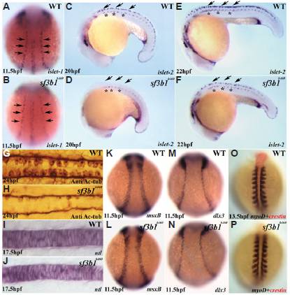
Fig. 8 Early development of ectodermal and mesodermal derivatives in sf3b1b460 mutants. (A,B) Whole-mount in situ hybridization with islet-1 antisense RNA probes shows that the number of Rohon-Beard precursor cells (arrows) is decreased in sf3b1b460mutants (dorsal view, anterior to top). (C-F) Whole-mount in situ hybridization with islet-2 antisense RNA probe shows deficiency in the development of Rohon-Beard sensory neurons (arrows), whereas the development of primary motor neurons is comparatively less affected in mutant embryos (asterisks). (G,H) Dorsal view of spinal cords labeled with anti-acetylated tubulin (Anti Ac-tub), anterior to left, showing that the Rohon-Beard sensory neuron phenotype persists at later stages in sf3b1b460 mutants. K-N: Induction of the neural plate border occurs normally in sf3b1b460 mutants. Whole-mount in situ hybridization with msxB (K,L) and dlx3 (M,N). (I,J;O,P) The derivatives from paraxial mesoderm and chordamesoderm are not affected in sf3b1b460mutants. (I,J) Lateral view of whole-mount in situ with ntl, anterior to left. (O,P) Dorsal view of double in situ with crestin (red) and myoD (blue), anterior to top.