
FIGURE
Fig. 5 - Supplemental 4
- ID
- ZDB-FIG-260507-17
- Publication
- Letelier et al., 2023 - Mutation of vsx genes in zebrafish highlights the robustness of the retinal specification network
- Other Figures
-
- Fig. 1
- Fig. 1 - Supplemental 1
- Fig. 1 - Supplemental 2
- Fig. 1 - Supplemental 3
- Fig. 2
- Fig. 2 - Supplemental 1
- Fig. 3
- Fig. 3 - Supplemental 1
- Fig. 4
- Fig. 4 - Supplemental 1
- Fig. 4 - Supplemental 2
- Fig. 4 - Supplemental 3
- Fig. 5
- Fig. 5 - Supplemental 1
- Fig. 5 - Supplemental 2
- Fig. 5 - Supplemental 3
- Fig. 5 - Supplemental 4
- text only
- All Figure Page
- Back to All Figure Page
Fig. 5 - Supplemental 4
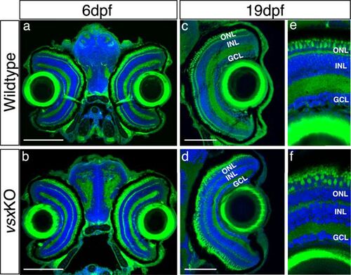
Normal eye size in vsxKO animals is observed at juvenile stages. (a-f). Visual system histological sections stained with nuclear marker DAPI and phalloidin-Alexa488 for actin filaments from wildtype (a, c and e, n=5 for both stages) and vsxKO (b, d and f) animals at 6dpf (a, b, n=8), and 19hpf (c, e, d and f, n=6). No changes in eye size are observed but lamination in vsx mutants is severely affected at later stages compared to siblings (e, f). dpf: days post fertilization, ONL: outer nuclear layer, INL: inner nuclear layer, GCL: ganglion cell layer. Scale bar (a-d): 100 µm. |
Expression Data
Expression Detail
Antibody Labeling
Phenotype Data
Phenotype Detail
Acknowledgments
This image is the copyrighted work of the attributed author or publisher, and
ZFIN has permission only to display this image to its users.
Additional permissions should be obtained from the applicable author or publisher of the image.
Full text @ Elife