
Fig. 4 - Supplemental 2
- ID
- ZDB-FIG-260507-11
- Publication
- Letelier et al., 2023 - Mutation of vsx genes in zebrafish highlights the robustness of the retinal specification network
- Other Figures
-
- Fig. 1
- Fig. 1 - Supplemental 1
- Fig. 1 - Supplemental 2
- Fig. 1 - Supplemental 3
- Fig. 2
- Fig. 2 - Supplemental 1
- Fig. 3
- Fig. 3 - Supplemental 1
- Fig. 4
- Fig. 4 - Supplemental 1
- Fig. 4 - Supplemental 2
- Fig. 4 - Supplemental 3
- Fig. 5
- Fig. 5 - Supplemental 1
- Fig. 5 - Supplemental 2
- Fig. 5 - Supplemental 3
- Fig. 5 - Supplemental 4
- text only
- All Figure Page
- Back to All Figure Page
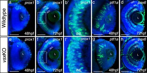
Analysis of INL markers prox1, ptf1a, and pax6 in WT and vsxKO retinas. (a-h). Confocal representative images of genes expressed in the INL by fluorescent in situ hybridization at different stages. At 48hpf, no obvious change in the expression of prox1 was detected between WT (a) and vsxKO (e) retinas. At 72hpf, the distribution of prox1 expression in the INL is affected in vsxKO (f and inset f’) compared to WT retinas (b and inset b’). No major changes in the expression of the amacrine cell markers ptf1a (c, g) at 48hpf and pax6 (d, h) at 72hpf were observed between WT (c, d) and vsxKO samples (g, h). ONL: outer nuclear layer, INL: inner nuclear layer, GCL: ganglion cell layer, L: lens, hpf: hours post-fertilization. Scale bar:50 µm. |