
FIGURE
Fig. 5 - Supplemental 3
- ID
- ZDB-FIG-260507-16
- Publication
- Letelier et al., 2023 - Mutation of vsx genes in zebrafish highlights the robustness of the retinal specification network
- Other Figures
-
- Fig. 1
- Fig. 1 - Supplemental 1
- Fig. 1 - Supplemental 2
- Fig. 1 - Supplemental 3
- Fig. 2
- Fig. 2 - Supplemental 1
- Fig. 3
- Fig. 3 - Supplemental 1
- Fig. 4
- Fig. 4 - Supplemental 1
- Fig. 4 - Supplemental 2
- Fig. 4 - Supplemental 3
- Fig. 5
- Fig. 5 - Supplemental 1
- Fig. 5 - Supplemental 2
- Fig. 5 - Supplemental 3
- Fig. 5 - Supplemental 4
- text only
- All Figure Page
- Back to All Figure Page
Fig. 5 - Supplemental 3
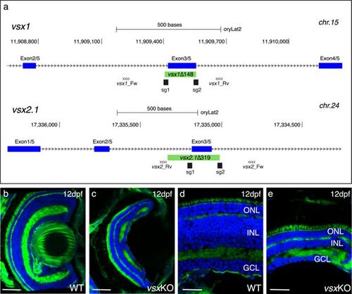
Mutation of vsx genes in medaka impairs INL differentiation and eye growth. (a) CRISPR/Cas9 was used to eliminate (green box) the DBD from vsx1 (top) and vsx2.1 (bottom) TFs in medaka. Blue boxes represent exons, black boxes the location of sgRNAs used and primers for screening are depicted as opposing arrowheads. (b-e). Histological sections from WT (b, d, n=4) and vsxKO central retinas (c, e, n=5) at 12dpf. ONL: outer nuclear layer, INL: inner nuclear layer, GCL: ganglion cell layer, hpf: hours post-fertilization, dpf: days post-fertilization. Scale bar (b-c): 50 µm, (d-e): 20 µm. |
Expression Data
Expression Detail
Antibody Labeling
Phenotype Data
Phenotype Detail
Acknowledgments
This image is the copyrighted work of the attributed author or publisher, and
ZFIN has permission only to display this image to its users.
Additional permissions should be obtained from the applicable author or publisher of the image.
Full text @ Elife