FIGURE

Figure 6.

ID
ZDB-FIG-260423-54
Publication
Wei et al., 2026 - A Novel CRYBB2 Splicing Mutation Is Associated With Lens Extracellular Matrix Remodeling and Vascular Alterations in Congenital Cataract
Other Figures
All Figure Page
Back to All Figure Page
Figure 6.

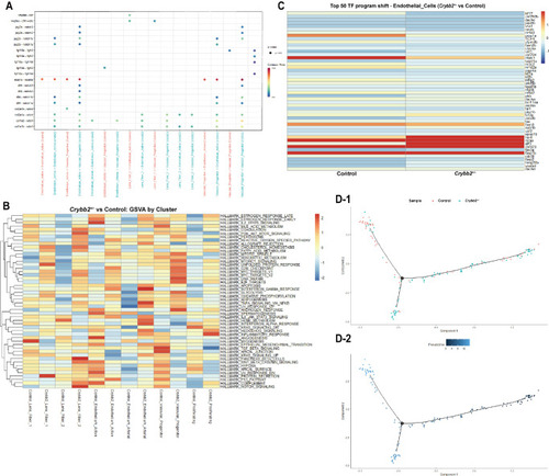
Transcription factor profiling and signaling pathway analysis in retinal vascular and lens-associated subclusters following crybb2 knockout. (A) Pseudotime trajectory of single-cell transcriptomes, colored by subcluster identity, showing developmental bifurcation in the Endothelium_Active population. (B) Pseudotime trajectory colored by experimental group, displaying distribution of WT and crybb2+/− knockout cells. (C) Heatmap of the top 50 transcription factors in endothelial cells. Rows represent TFs, and columns represent groups (WT vs. crybb2+/−). Expression values are scaled by row (scale = “row”). (D) BEAM branch heatmap of endothelial pseudotime trajectory, showing fate-biased gene modules identified by branch-dependent expression analysis. Genes are ordered by pseudotime and grouped by module identity.

Expression Data

Expression Detail
Antibody Labeling
Phenotype Data

Phenotype Detail
Acknowledgments
This image is the copyrighted work of the attributed author or publisher, and ZFIN has permission only to display this image to its users. Additional permissions should be obtained from the applicable author or publisher of the image. Full text @ Invest. Ophthalmol. Vis. Sci.