FIGURE

Figure 1.

ID
ZDB-FIG-260423-49
Publication
Wei et al., 2026 - A Novel CRYBB2 Splicing Mutation Is Associated With Lens Extracellular Matrix Remodeling and Vascular Alterations in Congenital Cataract
Other Figures
All Figure Page
Back to All Figure Page
Figure 1.

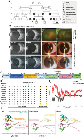
Clinical, genetic, and structural characterization of a five-generation family with CC. (A) Pedigree of a five-generation family with CC. Generations are labeled I–V. Symbols indicate sex and phenotype; the proband is marked with a black arrow. (B) Representative ophthalmic examinations of the proband (IV-6) and her affected parents (III-6 and III-7). The clinical imaging includes ophthalmic ultrasound examinations (for IV-6, III-6, and III-7), scanning laser ophthalmoscopy (for IV-6), and slit-lamp anterior segment photography (for III-6 and III-7). (C) Schematic diagram of CRYBB2 protein domains, with the splice-site mutation c.450-2A>G mapped to the intron-exon boundary. (D) Conservation analysis of the CRYBB2 locus across multiple species, illustrating cross-species sequence alignment. (E) AlphaFold-predicted three-dimensional structural models of WT and mutant CRYBB2 proteins, with Greek key motifs and terminal regions color-coded. Merged structural overlay is also shown. (F) Molecular dynamics simulations comparing WT and mutant CRYBB2 proteins, including RMSD trajectories.

Expression Data

Expression Detail
Antibody Labeling
Phenotype Data

Phenotype Detail
Acknowledgments
This image is the copyrighted work of the attributed author or publisher, and ZFIN has permission only to display this image to its users. Additional permissions should be obtained from the applicable author or publisher of the image. Full text @ Invest. Ophthalmol. Vis. Sci.